Artificial spine body support device for upper cervical spine

A technology of supporting device and vertebral body, which is applied to spinal implants and other directions, can solve the problems of high operation risk, poor shape matching, difficult operation, etc., and achieves high degree of humanization and human body structure, simple structure and convenient use. Effect

Pending Publication Date: 2018-08-17
GUANGZHOU GENERAL HOSPITAL OF GUANGZHOU MILITARY COMMAND +1
View PDF7 Cites 1 Cited by
  • Summary
  • Abstract
  • Description
  • Claims
  • Application Information

AI Technical Summary

Problems solved by technology

It is mainly reflected in: ①Surgical exposure is difficult. To remove the atlantoaxial lesion, the transoropharyngeal approach is generally required. If the lesion is large or the scope of reconstruction and fixation is large, it may be necessary to split the mandible and other traumatic methods. Large surgical approach, and this surgical exposure often requires the cooperation of stomatology doctors
②The surgical operation is risky. The spinal canal of the atlantoaxial contains the high cervical spinal cord and medulla oblongata. Removal of the lesion requires superb surgical skills. If there is a little carelessness, the injury to the medulla oblongata may lead to paralysis and death
[0005] At present, the general-purpose equipment used clinically is mainly commercialized titanium mesh. Ordinary titanium mesh is cylindrical in shape up and down. It is mainly designed for the lower cervical spine and thoracolumbar spine, which cannot meet the reconstruction needs of the complex anatomical shape of the atlantoaxial spine.
Although some scholars at home and abroad have improved implants such as titanium mesh and applied them to reconstruction after atlantoaxial resection, but the improved implants still have difficulties in fixation and poor shape matching when applied to this site. High fixation failure rate, low fusion rate and other defects, it is difficult to truly meet the needs of complex operations

Method used


Image

  • Artificial spine body support device for upper cervical spine
  • Artificial spine body support device for upper cervical spine
  • Artificial spine body support device for upper cervical spine

Examples


Embodiment

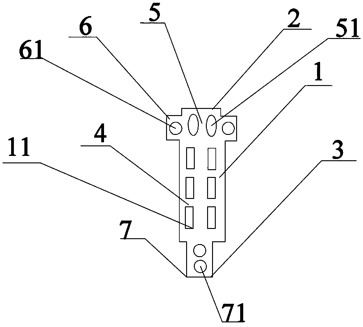
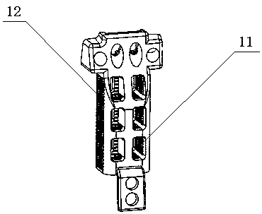
[0027] Such as Figure 1 to 3 Shown is an embodiment of an upper cervical artificial vertebra support device of the patent. The support device includes a hollow support member 1, a first support end surface 2, a second support end surface 3, a first support end surface 2, a second support end surface 3 is connected to both ends of the support 1 and forms a support body 4. The support device includes an occipital slope fixing member 5 for connecting the supporting body 4 with the occipital slope of the human body, an occipital condyle fixing member 6 for connecting the occipital condyle of the human body, and The lower vertebral body fixing member 7 connected to the lower vertebral body of the human body; the supporting member 1, the first supporting end surface 2, and the second supporting end surface 3 are provided with several penetration windows for infiltrating bone tissue; the first supporting surface 2 and the occiput The slope fixing member 5 and the occipital condyle fi...


PUM


Abstract

The invention relates to the technical field of medical devices, and in particular relates to an artificial spine body support device for the upper cervical spine. The support device comprises a support part with hollow inside, a first support end face and a second support end face, wherein the first support end face and the second support end face are connected to two ends of the support part, sothat a support main body is formed, the support device comprises an occipital bone slope fixing part used for connecting the support main body with the occipital bone slope of the human body, occipital bone condyle fixing parts used for connecting the support main body with the occipital bone condyles of the human body, and an inferior spine body fixing part used for connecting the support main body with the inferior spine body of the human body; a plurality of permeation windows used for permeation of the osseous tissue are arranged on the support part, the first support end face and the second support end face; the first support face is connected with the occipital bone slope fixing part and the occipital bone condyle fixing parts and is integrally formed with the occipital bone slope fixing part and the occipital bone condyle fixing parts, and the second support end face is in smooth connection with the inferior spine body fixing part. Through the arrangement, the artificial dentata support body suitable for the demands of the special anatomical structure of the craniocervical junctional area is designed, and a feasible solution is provided for the excision and reconstruction of the focus of the upper cervical spine performed by spine surgeons.

Description

Technical field [0001] This patent relates to the technical field of medical devices, and more specifically, to an upper cervical artificial vertebral body support device. Background technique [0002] Tuberculosis and tumors involving the upper cervical spine often severely damage the important structures of the atlantoaxial spine and compress the spinal cord, causing serious consequences of paralysis. Such patients need to surgically remove the lesion and reconstruct and fix the damaged structure to maintain the structural integrity and stability of the craniocervical junction and maintain normal physiological functions. [0003] Because the atlantoaxial vertebrae are located in the craniocervical junction area, their anatomical structure is special, the location is deep, and there are important nerves and blood vessels around, its resection and reconstruction operations are difficult points in spinal surgery. It is mainly reflected in: ①It is difficult to expose the atlantoaxia...

Claims


Application Information

Patent Timeline
Patent Type & Authority Applications(China)
IPC IPC(8): A61F2/44
CPCA61F2/44
Inventor 王建华张东升杨永强王迪王艺锰
Owner GUANGZHOU GENERAL HOSPITAL OF GUANGZHOU MILITARY COMMAND
Why Eureka
Try Eureka