Preparation method for tumour prosthesis of upper middle tibia

A prosthesis and tumor technology, applied in the direction of prosthesis, skull, bone implants, etc., can solve the problem that standardized prosthesis cannot meet personalized and precise treatment, the design process of individual custom prosthesis is complicated, and the quality of artificial joint is not as good as physiological Problems such as joints can be improved to improve the design process, strengthen long-term stability, and balance the overall mechanical distribution

Pending Publication Date: 2018-08-24
SOUTHERN MEDICAL UNIVERSITY
View PDF6 Cites 8 Cited by
  • Summary
  • Abstract
  • Description
  • Claims
  • Application Information

AI Technical Summary

Problems solved by technology

[0004] However, the quality of artificial joints currently used clinically is still inferior to that of physiological joints, and artificial prostheses still have deficiencies such as prosthetic infection, prosthetic loosening, and so on.
Clinically, joint-preserving tumor-type prostheses are scarce, and the basic principle of limb-

Method used


Image

  • Preparation method for tumour prosthesis of upper middle tibia
  • Preparation method for tumour prosthesis of upper middle tibia

Examples


Embodiment 1

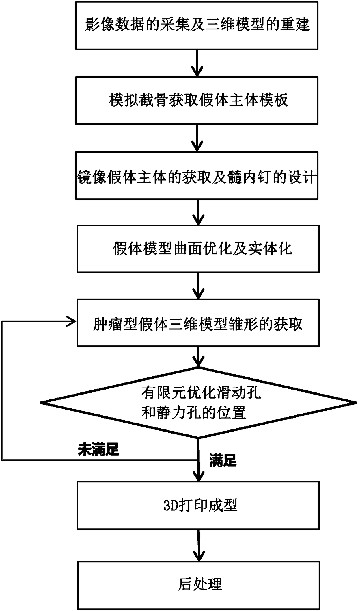
[0032] Such as figure 1 As shown, a method for preparing a tumor-type prosthesis in the middle and upper part of the tibia comprises the following steps in turn:

[0033] (1) Acquisition of image data and reconstruction of 3D model

[0034] Collect the computed tomography data of the bilateral tibia of the same target object, and import the obtained tomographic imaging data into 3D reconstruction software for image processing operations, and establish the 3D model of the bilateral tibia of the same target object and the 3D model of the bone tumor. The three-dimensional model of the bilateral tibia includes the three-dimensional model of the healthy tibia and the three-dimensional model of the affected tibia.

[0035] In this embodiment, the tomographic image data of the target tibia is acquired by a CT scanning imaging device. It should be noted that the tomographic image data can also be obtained by MRI scanning or micro-CT scanning imaging equipment; the 3D reconstruction ...

Embodiment 2

[0059] A method for preparing a tumor-type prosthesis in the middle and upper part of the tibia provided by this embodiment, other features are the same as in Embodiment 1, the difference is that the three-dimensional reconstruction software used in step (1) is 3D-doctor software, and the step (2) ) The three-dimensional modeling software used is Geomagic Studio software, and the computer-aided design software used in step (5) is UG NX software.

[0060] The method for preparing the tumor-type prosthesis in the middle and upper part of the tibia improves the design process of the traditional personalized tibial prosthesis, reduces the design difficulty and improves the efficiency; the tumor-type prosthesis prepared by this method not only retains the articular surface, Soft tissue reconstruction including the patellar ligament, medial and lateral collateral ligament, etc. is considered, and the position of the fixed plate on the articular surface avoids the soft tissue covering...


PUM


Abstract

The invention provides a preparation method for a tumour prosthesis of an upper middle tibia. The method comprises the steps that firstly, a three-dimensional reconstruction technology is adopted to reconstruct uninjured and affected side tibias, according to the tumor position, an osteotomy range is determined, a tibia prosthesis body is obtained through mirroring based on the uninjured side tibia, side wing steel plates and intramedullary nails which are arranged at two sides of sclerotin are fixed to two ends of the tibia prosthesis body through the computer aided design, and an STL model is exported for 3D printing and subsequent treatment. The preparation method is simple and quick and makes up for deficiency of an existing clinical tumour prosthesis of the upper middle tibia, based on the preparation method, the prepared prosthesis reserves the articular surface, the designed bone block fixing steel plate position of the articular surface keeps away from pes anserinus and other parts covered by nervus nerve soft tissues, the fixing is safer and more reliable, a far-end intramedullary nail adopts the combination technology of sliding holes and static holes, and the nail placing position is obtained through the finite element analysis technology, so that the overall mechanics distribution is more uniform after prosthesis implantation, and the long-term stability is reinforced.

Description

technical field [0001] The invention relates to the technical field of research and development of auxiliary devices for bone joint function reconstruction after bone tumor resection, in particular to a method for preparing a tumor-type prosthesis in the middle and upper tibia. Background technique [0002] The proximal tibia is the second most common site for primary malignant bone tumors after the distal femur. The anatomical structure of the proximal tibia is special, and it is closely related to the tibial nerve, common peroneal nerve, and blood vessels of the calf. The soft tissue coverage is weak, and it needs to be treated at the same time. For the tibiofibular joint, after resection of the tibial tuberosity, it is necessary to rebuild the knee extensor device, damage the starting point of the calf muscle, etc. It is difficult to salvage the limb, the complications are high, and the function of the knee joint is poor after reconstruction. Therefore, the extensive rese...

Claims


Application Information

Patent Timeline
IPC IPC(8): A61F2/28
CPCA61F2/28A61F2002/2892A61F2002/30952A61F2002/30948Y02P10/25
Inventor 黄文华谢普生张国栋
Owner SOUTHERN MEDICAL UNIVERSITY
Why Eureka
Try Eureka