Pulmonary vascular tree segmentation method with tubular structure enhancement and energy function combined

A tubular structure and energy function technology, which is applied in the field of medical image processing, can solve the problems of incomplete segmentation of small blood vessels, calculation amount of wrongly divided tracheal wall area, etc.

Inactive Publication Date: 2018-09-04
UNIV OF SHANGHAI FOR SCI & TECH
View PDF3 Cites 12 Cited by
  • Summary
  • Abstract
  • Description
  • Claims
  • Application Information

AI Technical Summary

Problems solved by technology

[0003] The present invention aims at the problems of incomplete segmentation of small blood vessels, misclassification of tracheal wall area and large amount of calculation in the segmentation of pulmonary vascular tree, and proposes a pulmonary vascular tree segmentation method combining tubular structure enhancement and energy function, To achieve effective segmentation of the pulmonary vascular tree

Method used


Image

  • Pulmonary vascular tree segmentation method with tubular structure enhancement and energy function combined
  • Pulmonary vascular tree segmentation method with tubular structure enhancement and energy function combined
  • Pulmonary vascular tree segmentation method with tubular structure enhancement and energy function combined

Examples


Embodiment Construction

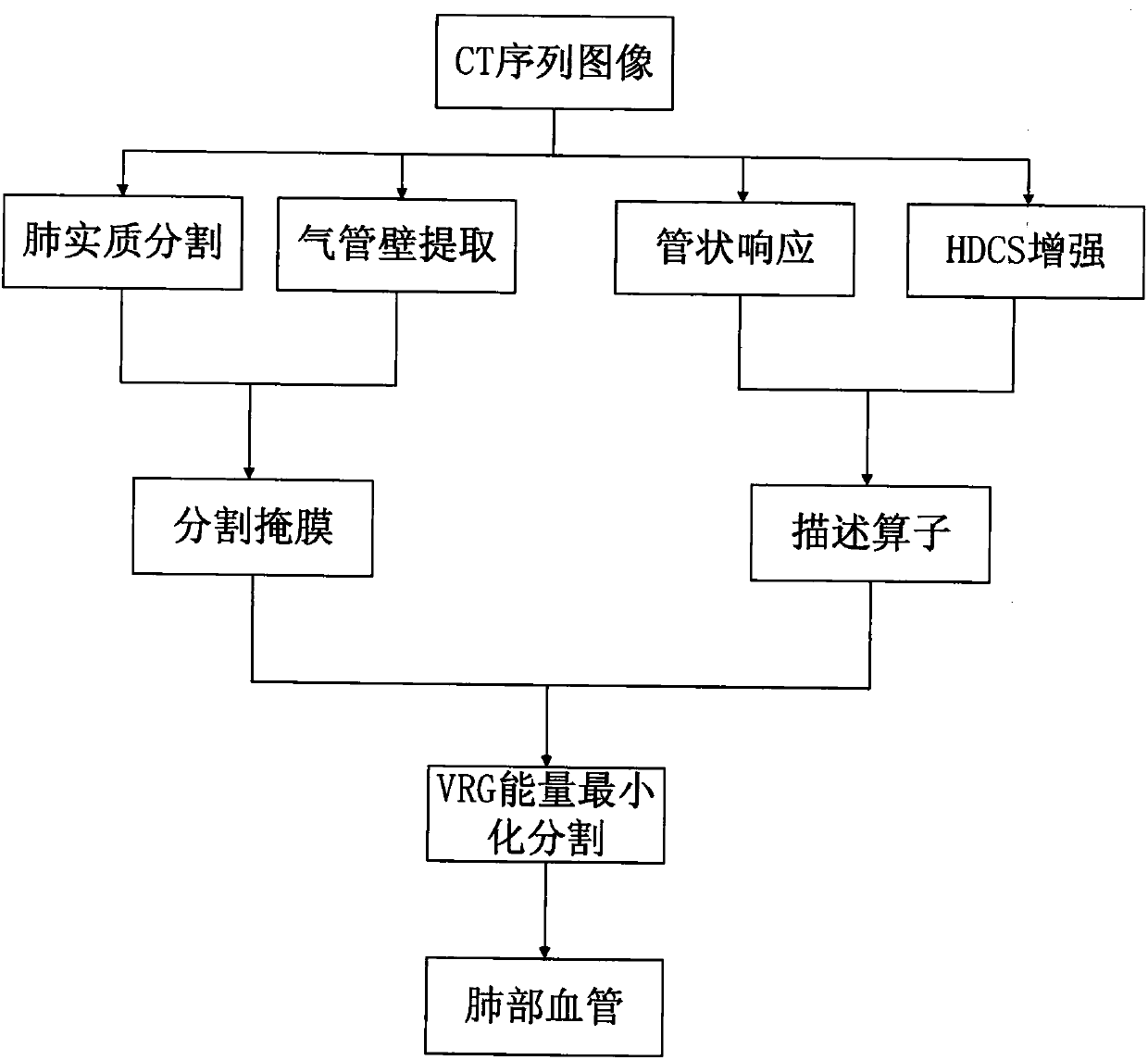
[0080] The segmentation method of pulmonary vascular tree combining tubular structure enhancement and energy function uses Pock function to calculate the responsivity of tubular structure to detect potential vascular regions. Then the original image is enhanced by a tubular structure enhancement algorithm based on diffusion tensor, which reduces the influence of noise on the original image and enhances the vessel area. Finally, the calculation result of Pock function and the result of image enhancement are combined to construct a region description operator, and the pulmonary blood vessels are finely segmented using the VRG method, which is a minimum energy segmentation method.

[0081] like figure 1 The flow chart of the pulmonary vessel tree segmentation method combined with tubular structure enhancement and energy function is shown, including the following steps:

[0082] Step 1, input the tomographic image (original image) of chest CT sequence in DICOM format to be segmen...


PUM


Abstract

The invention relates to a pulmonary vascular tree segmentation method with tubular structure enhancement and an energy function combined. A Pock function is used to calculate the response of a tubular structure, and a potential vascular region is thus detected; a tubular structure enhancement algorithm based on a diffusion tensor is then adopted to enhance the original image, influences on the original image by noise are reduced, and the vascular region is enhanced; and finally, a Pock function calculation result and an image enhancement result are combined to build a region description operator, and a minimum energy segmentation method VRG method is used to finely segment the pulmonary vessel. The segmentation result shows that while the method segments a main pulmonary vessel, a large amount of tiny vessels are also extracted, and the segmentation result is little influenced by the noise. The method is high in specificity, the sensitivity is strong, a vessel and a tracheal wall areacan be distinguished, and the segmentation result accuracy is further improved.

Description

technical field [0001] The invention relates to a medical image processing technology, in particular to a pulmonary blood vessel tree segmentation method combining tubular structure enhancement and energy function. Background technique [0002] Pulmonary blood vessels are composed of pulmonary arteries and pulmonary veins, and are one of the most complex vascular structures in various tissues and organs of the human body. Starting from the pulmonary aorta and main pulmonary vein, the pulmonary blood vessels branch out step by step to form a tree-like vascular tree structure. In clinical diagnosis, accurate acquisition of the anatomical structure information of the pulmonary vascular tree is an important reference for assessing the risk of pulmonary hypertension and the basis for automatic detection of pulmonary embolism. It is also beneficial to reduce the false positive rate of pulmonary nodule detection. In clinical research, effectively separating the pulmonary vascular ...

Claims


Application Information

Patent Timeline
Patent Type & Authority Applications(China)
IPC IPC(8): G06T7/11G06T7/155G06T5/00
CPCG06T5/002G06T2207/30061G06T2207/30101G06T7/11G06T7/155
Inventor 段辉宏聂生东王丽嘉龚敬
Owner UNIV OF SHANGHAI FOR SCI & TECH
Why Eureka
Try Eureka