Method and system for melanoma image tissue segmentation based on deep neural network

A deep neural network, melanoma technology, applied in the field of melanoma image tissue segmentation

Active Publication Date: 2018-09-07
SOUTH CHINA UNIV OF TECH
View PDF2 Cites 26 Cited by
  • Summary
  • Abstract
  • Description
  • Claims
  • Application Information

AI Technical Summary

Problems solved by technology

The main limitation is that these methods achieve very limited results

Method used


Image

  • Method and system for melanoma image tissue segmentation based on deep neural network
  • Method and system for melanoma image tissue segmentation based on deep neural network
  • Method and system for melanoma image tissue segmentation based on deep neural network

Examples


Embodiment Construction

[0055] The present invention will be further described below in conjunction with specific examples.

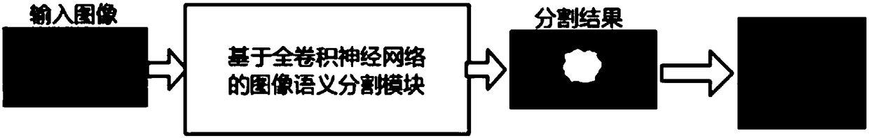
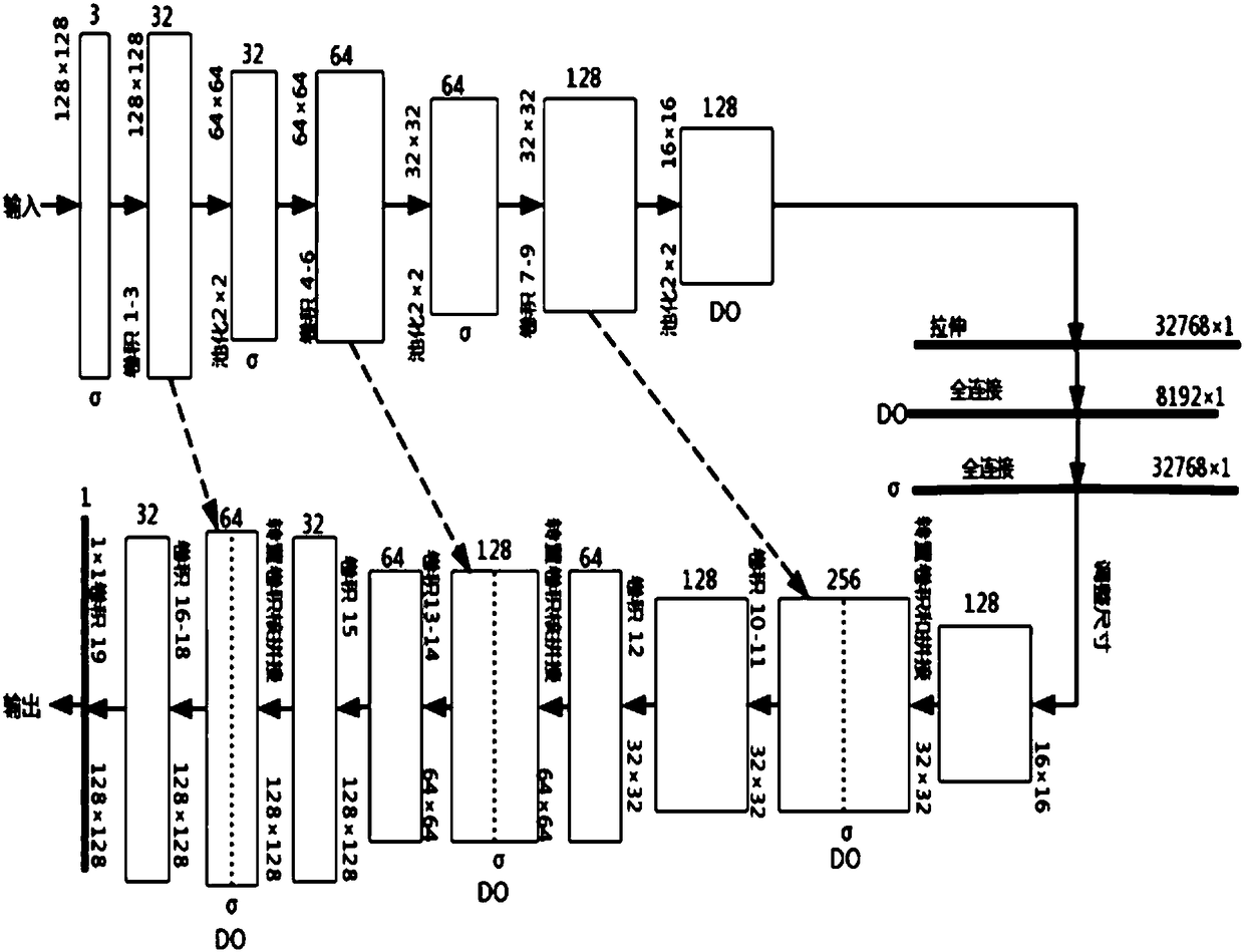
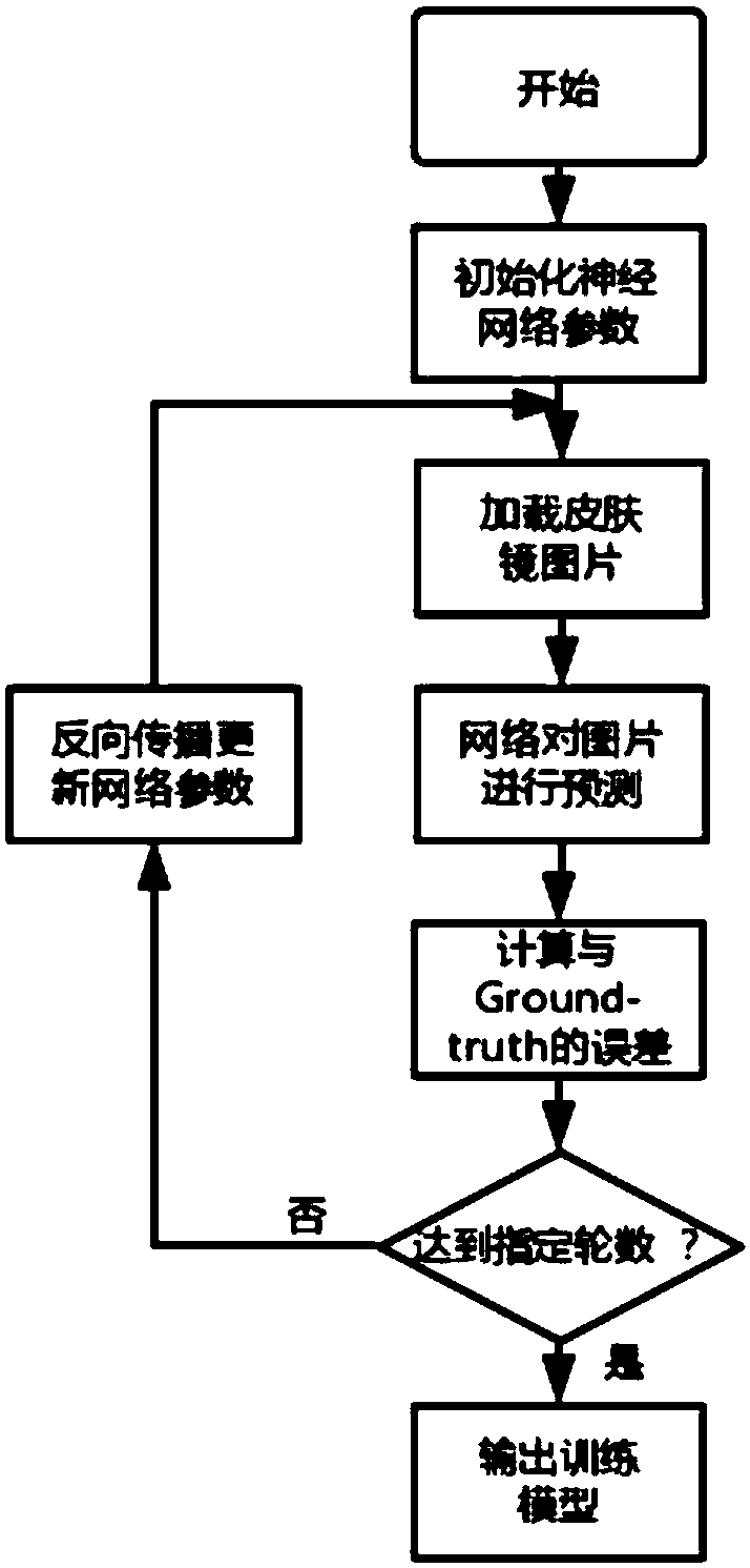
[0056] This embodiment provides a method for tissue segmentation of melanoma pictures based on a deep neural network, which is an improved implementation of a neural network for image semantic segmentation, and its network structure is as follows figure 2 shown; the method first trains the neural network model with existing dermoscopic pictures about melanoma, and selects the model with the best training effect as the final model. The role of the model is to mark the skin lesion area in the picture; the input of the model is a preprocessed melanoma dermoscopy picture, and the output is a segmented picture with only black and white colors, and white represents suspicious skin lesions Areas, black represent normal skin tissue. The method is carried out as follows:

[0057] Step 1, build deep neural network model, this network is a 21-layer deep neural network, the structure o...


PUM


Abstract

The present invention discloses a method and a system for melanoma image tissue segmentation based on a deep neural network. A deep neural network technology is employed to solve the tissue segmentation problem of dermatoscope images of melanoma, and the method and the system for melanoma image tissue segmentation based on a deep neural network are mainly a medical image analysis processing technology. An improved deep neural network structure to perform modeling, dermatoscope images having segmentation tags are employed to train the model, and the trained model has an ability of suspicious tissues to new dermatoscope images. The method and the system are to locate suspicious areas in the melanoma dermatoscope images and to perform pixel segmentation. The method and the system for melanomaimage tissue segmentation based on the deep neural network employ a new deep learning technology, fully develop the capacity of collection of various hierarchical features of the image data, can be applied to the modeling process, can perform location and segmentation of the suspicious skin tissues and can provide good reference for further analysis by dermatologists.

Description

technical field [0001] The present invention relates to the technical field of medical image processing, in particular to a method and system for tissue segmentation of melanoma images based on a deep neural network. Background technique [0002] With the development of computer technology, medical imaging technology and the increasing social medical needs year by year, medical data, especially medical image data, is increasing at an unprecedented rate. Accurate identification of medical images has obvious social significance and use value. At the same time, in the field of computer vision and pattern recognition, the supervised learning method in deep learning is becoming a hot spot in theory and industry, and breakthroughs have been made. In view of the fact that most of the traditional medical image analysis uses manual annotation or artificial extraction of features and then uses machine learning, it is not only time-consuming and laborious, difficult to extract, but als...

Claims


Application Information

Patent Timeline
Patent Type & Authority Applications(China)
IPC IPC(8): G06T7/11
CPCG06T7/11G06T2207/20081G06T2207/20084G06T2207/30088
Inventor 柏朋成赵跃龙张声超
Owner SOUTH CHINA UNIV OF TECH
Why Eureka
Try Eureka