
Automatic segmentation method and system of esophageal endoscopic OCT image hierarchy structure

An automatic segmentation and hierarchical structure technology, applied in the field of medical image processing, can solve problems such as insufficient segmentation accuracy, unfavorable automatic segmentation, and difficulty in setting attenuation function parameters, and achieve the effect of improving accuracy

Active Publication Date: 2018-11-06
SUZHOU UNIV
View PDF6 Cites 8 Cited by
  • Summary
  • Abstract
  • Description
  • Claims
  • Application Information

AI Technical Summary

Problems solved by technology

However, this method does not take into account the influence of interstitial fluid, abnormal structure and probe membrane in the esophageal OCT image, so it is often unable to give a stable segmentation result; in addition, the parameter setting of the attenuation function in the second step of the method is difficult, It is not conducive to the realization of automatic segmentation; finally, because only the vertical gradient is used as the weight of graph search, it is slightly insufficient in segmentation accuracy

Method used


Image

  • Automatic segmentation method and system of esophageal endoscopic OCT image hierarchy structure
  • Automatic segmentation method and system of esophageal endoscopic OCT image hierarchy structure
  • Automatic segmentation method and system of esophageal endoscopic OCT image hierarchy structure

Examples


Embodiment Construction

[0068] Exemplary embodiments of the present disclosure will be described in more detail below with reference to the accompanying drawings. Although exemplary embodiments of the present disclosure are shown in the drawings, it should be understood that the present disclosure may be embodied in various forms and should not be limited by the embodiments set forth herein. Rather, these embodiments are provided for more thorough understanding of the present disclosure and to fully convey the scope of the present disclosure to those skilled in the art.

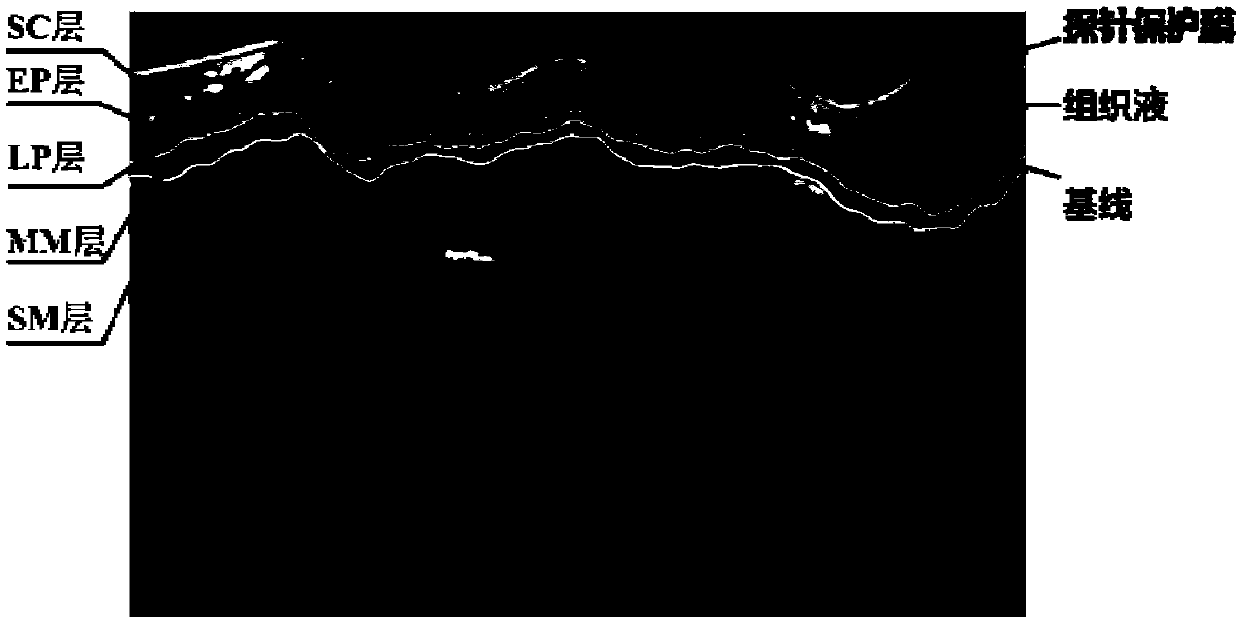
[0069] According to an embodiment of the present invention, an automatic segmentation method for the hierarchical structure of esophageal endoscopic OCT images is proposed. The collected OCT images and structures of the esophagus are as follows: figure 1 As shown, a total of 6 layer boundaries corresponding to 5 tissue layers need to be segmented. Muscularis Mucosae (MM) and Submucosa (SM). The specific segmentation is based on t...


PUM


Abstract

The invention discloses an automatic segmentation method and a system of an esophageal endoscopic OCT image hierarchy structure. The method comprises the following steps: image preprocessing, whereinan image is filtered and searched, and pixel values are complemented; image planarization, wherein the whole esophageal tissue in the image is straightened and corrected; segmentation of an image organization layer, wherein a current segmentation layer is used as a reference and the searching area of a next layer to be segmented is obtained in a mode of translating certain pixels downward and segmentation layer by layer is performed; image unplanarization, wherein the segmented image is inversely translated to obtain the final segmentation result. The invention provides a scheme for respondingto interference of a probe protective film, tissue liquid and the like, so as to avoid subsequent influences on tissue layer segmentation. The invention combines the Canny edge detection with the gradient operator, and proposes a new image segmentation method weight design scheme. The scheme uses the Canny operator to improve the precision of edge detection, and uses the gradient operator to compensate the missing edge in the Canny operator. The accuracy of segmentation is improved by combining the two operators.

Description

technical field [0001] The invention relates to the field of medical image processing, in particular to an automatic segmentation method and system for an esophageal endoscopic OCT image hierarchy. Background technique [0002] OCT technology can realize rapid and non-destructive high-resolution imaging of living bodies. It has become a technical hotspot in the field of medical imaging in recent years and has been widely used in clinical medicine. Endoscopic OCT technology is an important branch of OCT. Through the use of endoscopes, it can enter the internal cavity of the human body under low-invasive conditions and perform real-time imaging of biological living tissues, providing a new direction for clinical diagnosis. The diagnosis of esophageal diseases is an important clinical application of endoscopic OCT technology. The esophageal tissue under the endoscope shows a multi-layer structure, but the OCT technology itself will cause a lot of speckle noise in the imaging, ...

Claims


Application Information

Patent Timeline
Patent Type & Authority Applications(China)
IPC IPC(8): G06T7/00G06T7/12
CPCG06T7/0012G06T7/12G06T2207/10101G06T2207/30004
Inventor 王丽荣王聪甘萌
Owner SUZHOU UNIV
Learn More