

A new way to diagnose and monitor oral cancer
A technology for oral cancer and oral leukoplakia, which is applied in the new field of diagnosis and monitoring of oral cancer, and can solve the problems of traumatic and irritating lesions, affecting observation, and limiting inspection repeatability
- Summary
- Abstract
- Description
- Claims
- Application Information
AI Technical Summary
Problems solved by technology
Method used
Image

Examples
Embodiment 1
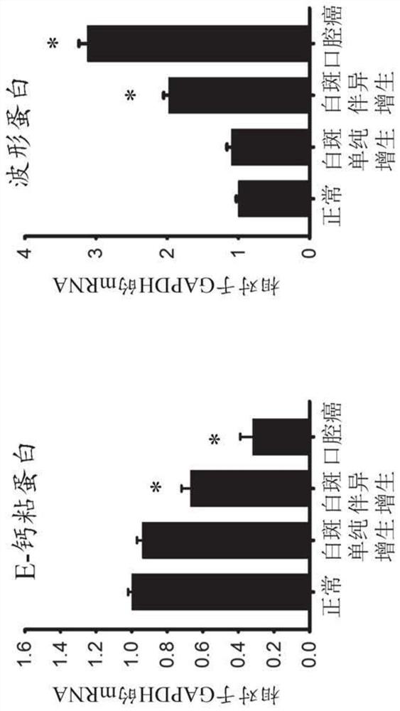
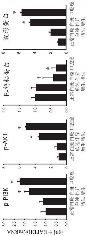
[0073] Example 1 Changes of important signal pathways during oral cancer before cancer
[0074] method
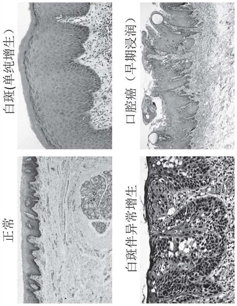
[0075] Choose clinical and pathological diagnosis as tissue specimens of oral mucosalous whiteprint (simple hyperplasia), white spots with abnormal hyperplasia and leukular cancer (oral squamous cell carcinoma) as the study object. Exemplary H & E Dyeing pathology of the tissue specimen figure 1 Indicated.
[0076] Based on pathological diagnosis results: divided into a white spot simple hyperplasia group (n = 15); white spots with abnormal hyperplasia group (n = 10), cancer group, also known as oral cancer group (n = 15).
[0077] Normal control group (N = 5) tissue specimens were selected from the group consolidation of oral mucosa disease, and some normal tissues needed for surgical treatment and willing to provide patients for research.
[0078] Immunohistochemical detection
[0079] The tissue samples of normal group, white spots, abnormal hyperplasia, and oral cancer gro...
Embodiment 2
[0094] Example 2 Oral saliva extracted body carry miR-185, consistency, reactive oral cancer pre-lesion
[0095] method
[0096] Subject: Patients with oral mucosa in the clinical and pathological diagnosis of the above Example 1, patients with oral mucosa, abnormal hyperplasia, oral cancer (oral squamous cell carcinoma) and normal human oral The following methods were collected, and the oral subordinate bodies were purified.
[0097] The patient or normal people did not have a mouth before taking a varying, and the water was fasted for 1 hour. Sitting in saliva, the head is naturally low, and the saliva in the mouth is naturally spit out of the disposable tray, about 2 ml, do not cough. The collected saliva is immediately placed in a small center tube, stored at 4 ° C.
[0098] The sample 4 ° C, 410,000 × g was centrifuged for 20 minutes, and the supernatant was filtered twice by flash of 0.22 μm, and the ultra-speed centrifuge (Beckman Coulter, Brea, Ca, US) was filtered by a T...
Embodiment 3
[0113] Example 3 Dynamic changes in oral saliva extract in the transformation of oral cancer
[0114] method
[0115] The purified saliva extremum was collected in the above Example 2, quantitative analysis was carried out by the following method.
[0116] Purified saliva extracrystology samples evaluated the size and frequency of extracted in Malverninstruments, WESTBORANOUGH (MA).
[0117] The nanoparticle tracking analysis was performed by using the Nanosight NS300 instrument (Nanosight NTA 2.3 NanopArticle Tracking Andand Analysis Release Version Build 0025). The size distribution of the preparation of the preparation is analyzed by using the Nanosight LM10 system (Nanosight, Wiltshire, United Kingdom) equipped with fast video capture and particle tracking software, and quantitatively analyzes the size of the preparation of the Brown. The purified exosomass was diluted in 400 μl of 1 × PBS / 5 mM EDTA solution, and then the sample was injected into the Nanosight sample chambe...
PUM
| Property | Measurement | Unit |
|---|---|---|
| diameter | aaaaa | aaaaa |
Abstract
Description
Claims
Application Information