High-frequency ultrasonic phased array for ophthalmology and preparation method thereof

An ultrasonic phased array and ultrasonic phased control technology, which is used in eye testing equipment, eye examination, blood flow measurement devices, etc. The effect of blood flow condition, improved resolution ability, good beam deflection ability

Inactive Publication Date: 2018-11-30
HUAZHONG UNIV OF SCI & TECH
View PDF14 Cites 6 Cited by
  • Summary
  • Abstract
  • Description
  • Claims
  • Application Information

AI Technical Summary

Problems solved by technology

[0005] In view of the above defects or improvement needs of the prior art, the present invention provides a high-frequency ultrasonic phased array array for ophthalmology and its preparation method, thereby solving the problems of the prior art with limited imaging resolution and inability to monitor the fundus in real time blood flow condition

Method used


Image

  • High-frequency ultrasonic phased array for ophthalmology and preparation method thereof

Examples


preparation example Construction

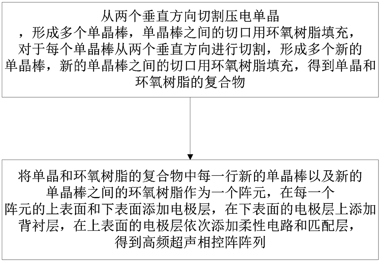
[0023] like figure 1 Shown, a kind of preparation method of the high-frequency ultrasonic phased array array that is used in ophthalmology, comprises:

[0024] (1) Utilize dry etching or laser cutting to cut the piezoelectric single crystal from two vertical directions to form a plurality of single crystal rods, the incisions between the single crystal rods are filled with epoxy resin, and the gaps between the single crystal rods The center distance is 70 μm-140 μm, and the width of the slit between the single crystal rods is 10 μm-15 μm; after the epoxy resin is cured, the air bubbles in the slit between the single crystal rods are removed in vacuum, and then for each single crystal The rods are cut from two vertical directions by dry etching or laser cutting to form multiple new single crystal rods, the cuts between the new single crystal rods are filled with epoxy resin, and the gaps between the new single crystal rods are filled with epoxy resin. The distance between the ...

Embodiment 1

[0027] A method for preparing a high-frequency ultrasonic phased array for ophthalmology, comprising:

[0028] (1) Utilize dry etching to cut PIN-PMN-PT from two perpendicular directions, form a plurality of PIN-PMN-PT sticks, the incision between PIN-PMN-PT sticks is filled with epoxy resin, described PIN- The center distance between the PMN-PT rods is 70 μm, and the width of the incision between the PIN-PMN-PT rods is 10 μm; when the epoxy resin is cured, remove the incision between the PIN-PMN-PT rods in vacuum The air bubbles, and then use dry etching to cut from two perpendicular directions for each PIN-PMN-PT rod, forming multiple new PIN-PMN-PT rods, and the cuts between the new PIN-PMN-PT rods Fill with epoxy resin, the center distance between the new PIN-PMN-PT rods is 35 μm, the width of the slit between the new PIN-PMN-PT rods is 10 μm, when the epoxy resin is cured, Remove air bubbles from the cut between the fresh PIN-PMN-PT rods in vacuo to obtain a 1-3 composit...

Embodiment 2

[0031] A method for preparing a high-frequency ultrasonic phased array for ophthalmology, comprising:

[0032] (1) Utilize laser cutting to cut PMN-PT from two vertical directions, form a plurality of PMN-PT rods, the otch between PMN-PT rods is filled with epoxy resin, and the center distance between described PMN-PT rods is 140 μm, the width of the kerf between the PMN-PT rods is 15 μm; when the epoxy resin is cured, remove the air bubbles of the kerf between the PMN-PT rods in vacuum, and then use a dry method for each PMN-PT rod Etching cuts from two perpendicular directions, forms a plurality of new PMN-PT rods, the incision between the new PMN-PT rods is filled with epoxy resin, and the center distance between the new PMN-PT rods is 70 μm, the width of the slit between the new PMN-PT rods is 15 μm, after the epoxy resin is cured, remove the air bubbles in the slit between the new PMN-PT rods in vacuum to obtain PMN-PT and epoxy 1-3 complexes of resins.

[0033](2) In t...


PUM


Abstract

The invention discloses a high-frequency ultrasonic phased array for ophthalmology and a preparation method thereof, wherein the preparation method includes: cutting piezoelectric monocrystal from twovertical directions to form multiple monocrystal rods, filling cuts of the monocrystal rods with epoxy resin, cutting each monocrystal rod from two vertical directions to form a plurality of new monocrystal rods, and filling cuts of the new monocrystal rods with epoxy resin to obtain monocrystal-epoxy resin complex; using the new monocrystal rods of each row in the monocrystal-epoxy resin complexand the epoxy resin between the new monocrystal rods as an array element, adding electrode layers to upper and lower surfaces of each array element, adding a backing layer to an electrode layer of each lower surface, and adding a flexible circuit and a matching layer to the electrode layer of each upper surface so that the high-frequency ultrasonic phased array is obtained. The high-frequency ultrasonic phased array has high resolving power for imaging and can monitor fundal blood flow condition in real time.

Description

technical field [0001] The invention belongs to the field of ultrasonic transducer imaging, and more specifically relates to a high-frequency ultrasonic phased array used in ophthalmology and a preparation method thereof. Background technique [0002] Ultrasound imaging technology plays an important role in ophthalmic imaging due to its safety, real-time and convenient features. In general, ultrasonic transducers for medical imaging are mainly divided into two categories: single-element transducers and array transducers (including linear arrays and phased arrays). Single-element transducers rely on mechanical scanning for acoustic imaging, and the best image quality can only be achieved at the focal point of the transducer. Compared with single-element transducers, array transducers have higher stability in acoustic image scanning. Therefore, ultrasound transducer arrays have several advantages over ultrasound transducer units, such as real-time measurement of blood flow, ...

Claims


Application Information

Patent Timeline
Patent Type & Authority Applications(China)
IPC IPC(8): A61B8/10A61B8/06
CPCA61B8/06A61B8/10
Inventor 朱本鹏雷爽张桃欧阳君杨晓非
Owner HUAZHONG UNIV OF SCI & TECH
Why Eureka
Try Eureka