Method for automatically segmenting head and neck tumors in MRI image

A head and neck tumor, in-image technology, applied in the field of medical images, can solve the problems of reducing segmentation accuracy and robustness, and achieve the effect of good segmentation performance

Active Publication Date: 2018-12-07
SICHUAN UNIV
View PDF6 Cites 8 Cited by
  • Summary
  • Abstract
  • Description
  • Claims
  • Application Information

AI Technical Summary

Problems solved by technology

However, the accuracy and robustness of segmentation is reduced due to the labor-intensive nature of manual segmentation and disagreement between different radiologists

Method used


Image

  • Method for automatically segmenting head and neck tumors in MRI image
  • Method for automatically segmenting head and neck tumors in MRI image
  • Method for automatically segmenting head and neck tumors in MRI image

Examples


Embodiment Construction

[0035] In order to make the purpose, technical solution and advantages of the present invention clearer, the present invention will be further elaborated below in conjunction with the accompanying drawings.

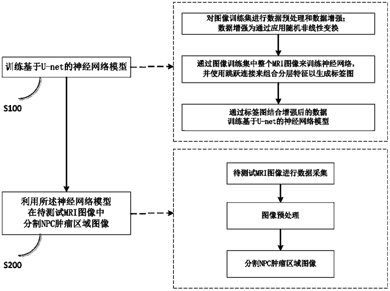
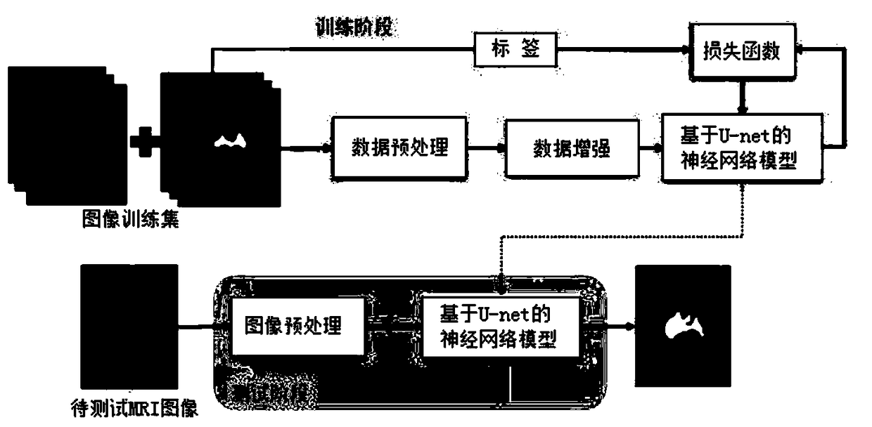
[0036] In this example, see figure 1 and figure 2 As shown, the present invention proposes an automatic head and neck tumor segmentation method in an MRI image, comprising steps:

[0037] S100, training a neural network model based on U-net: the neural network model includes a contraction encoder for analyzing input MRI images and an extended decoder for generating label map output; using skip connections in the U-net architecture will shallow The appearance feature representation of the encoding layer is combined with the high-level feature representation of the depth decoding layer;

[0038] Train the neural network model based on U-net, comprising steps:

[0039] S101, performing data preprocessing and data enhancement on the image training set; data enhancement is t...


PUM


Abstract

The invention discloses a method for automatically segmenting head and neck tumors in an MRI image, comprising the steps of training a neural network model based on a U-net, wherein the neural networkmodel comprises a contractive encoder for analyzing an input MRI image and an extension decoder for generating a label map output; combining the appearance feature representation of a shallow encoding layer with the advanced feature representation of a depth decoding layer by using a jump connection in the U-net architecture; and using the neural network model to segment a NPC tumor region imagein the MRI image to be tested. The method can rapidly, stably accurately and automatically segment the NPC tumors in the MRI image.

Description

technical field [0001] The invention belongs to the technical field of medical images, in particular to an automatic head and neck tumor segmentation method in MRI images. Background technique [0002] Among head and neck tumors, nasopharyngeal carcinoma (NPC) is the most common type causing high mortality; most NPC patients have missed the best treatment period before diagnosis of NPC. Accurate tumor delineation in magnetic resonance imaging (MRI) images plays a crucial role in guiding radiation therapy. [0003] Therefore, early diagnosis of NPC is particularly important in clinical applications. NPC patients are usually diagnosed based on manual segmentation and medical image analysis. Compared with other types of tumors such as brain tumors and lung tumors, NPC tumors have a more complex anatomy and often have similar intensities to surrounding tissues such as brainstem, cochlea, parotid gland, and lymph; additionally, tumors from different NPC patients Usually exhibi...

Claims


Application Information

Patent Timeline
Patent Type & Authority Applications(China)
IPC IPC(8): G06T7/11
CPCG06T2207/10088G06T2207/20081G06T2207/20084G06T2207/30096G06T7/11
Inventor 王艳何坤林峰吴锡周激流
Owner SICHUAN UNIV
Why Eureka
Try Eureka