Method for fine segmentation of eye ground optic disc based on SLIC super-pixel segmentation

A superpixel segmentation and fine segmentation technology, applied in image analysis, image enhancement, image data processing and other directions, can solve the problems of long time, high computational complexity and short time consumption.

Active Publication Date: 2018-12-07
UNIV OF ELECTRONICS SCI & TECH OF CHINA
View PDF9 Cites 15 Cited by
  • Summary
  • Abstract
  • Description
  • Claims
  • Application Information

AI Technical Summary

Problems solved by technology

[0007] The purpose of the present invention is to solve the problem that the current method for finely segmenting the optic disc of the fundus image depends on the huge number of samples or the initial contour setting, the calculation complexity is high, the time-consuming is long and the segmentation is not fine. The fundus optic disc fine segmentation method of pixel segmentation r

Method used


Image

  • Method for fine segmentation of eye ground optic disc based on SLIC super-pixel segmentation
  • Method for fine segmentation of eye ground optic disc based on SLIC super-pixel segmentation
  • Method for fine segmentation of eye ground optic disc based on SLIC super-pixel segmentation

Examples


Embodiment 1

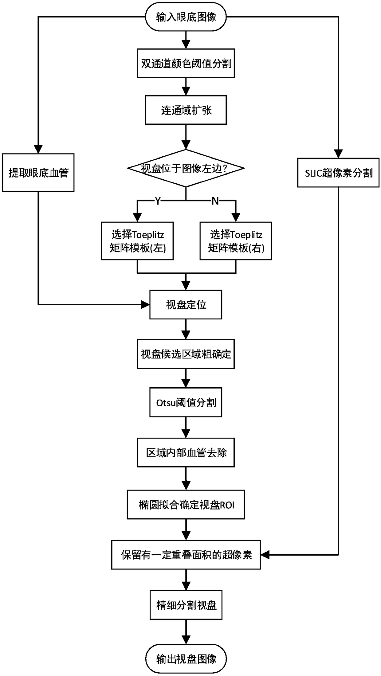
[0141] A fine segmentation method of fundus optic disc based on SLIC superpixel segmentation provided by a preferred embodiment of the present invention, the flow chart is as follows figure 1 As shown, the method steps include:

[0142] Step 1. Perform SLIC superpixel segmentation, fundus blood vessel image segmentation, and R, G dual-channel color threshold segmentation on the input fundus image, extract connected domains from the image after color threshold segmentation, expand the connected domains, and then count the fundus images Number of pixels on either side of the vertical centerline.

[0143] The SLIC superpixel segmentation step in the step 1 is specifically:

[0144] Step 1.1.11, for the input fundus image G, first convert the image into a five-dimensional feature vector V=[l, a, b, x, y], where [l, a, b] is the pixel color and belongs to the CIELAB color space , [x, y] is the pixel position.

[0145] Step 1.1.12, due to the different measurement methods of colo...


PUM


Abstract

The invention discloses a method for fine segmentation of the eye ground optic disc based on SLIC super-pixel segmentation. The steps are as follows: super-pixel segmentation, vascular segmentation based on morphological processing and R-G double-channel color threshold segmentation are carried out on an input eye ground image, and after expansion of a connected region obtained from color threshold segmentation, a corresponding Toeplitz matrix template is selected according to the pixel coordinates of the connected region to filter an eye ground vascular image to obtain the center position ofthe optic disc; and then, a candidate area of the optic disc is extracted and the internal blood vessels are removed, threshold segmentation is carried out on the candidate area of the optic disc through a binarization method, the ROI (Region of Interest) of the optic disc ellipse is determined by using an ellipse fitting method based on least squares, super pixels with a certain overlapping areaare retained according to the SLIC super-pixel segmentation result, and thus, fine segmentation of the optic disc is completed. Through the method, automatic positioning and fine segmentation of the optic disc are realized, better contour information of the optic disc can be retained, less time is consumed, other follow-up processing of the eye ground image is facilitated, and assistant diagnosiscan be provided for ophthalmologists.

Description

technical field [0001] The invention belongs to the technical field of segmentation methods for optic discs of fundus images, and in particular relates to a method for finely segmenting optic discs of fundus based on SLIC superpixel segmentation. Background technique [0002] The optic disc, or optic disc, is considered to be one of the main features of retinal fundus images. Optic disc detection is a key preprocessing part of many algorithms designed for automatic extraction of retinal anatomy and lesions, and a related module of most retinal lesion screening systems. The optic disc is often used as a marker for other fundus features, such as the fairly constant distance between the optic disc and the fovea, which can be used as a priori knowledge to help estimate the position of the macula. In addition, changes in the shape, color, or depth of the optic disc are indicators of various ophthalmic pathologies, especially glaucoma, so the dimensionality of the optic disc is o...

Claims


Application Information

Patent Timeline
IPC IPC(8): G06T7/12G06T7/33
CPCG06T2207/30041G06T7/12G06T7/33
Inventor 彭真明王慧周宇王卓然赵学功何艳敏袁国慧曲超范文澜尹利贺晨
Owner UNIV OF ELECTRONICS SCI & TECH OF CHINA
Why Eureka
Try Eureka