
Hemostatic bag for obstetrics and gynecology department

A technology of obstetrics and gynecology and capsules, which is applied in the field of medical devices, can solve the problems of reducing the hemostatic function of drugs, polluting drugs, leaking hospital beds, etc., and achieve the effects of convenient post-processing, simple export control, and clear export control

Inactive Publication Date: 2018-12-11
武汉半边天医疗技术发展有限公司
View PDF8 Cites 4 Cited by
  • Summary
  • Abstract
  • Description
  • Claims
  • Application Information

AI Technical Summary

Problems solved by technology

But it has the following disadvantages: during the expansion process of the uterine cavity capsule, the dirt remaining in the uterine cavity is pushed to the upper and lower ends of the uterine cavity, and is exported through two drainage tubes respectively. Although the dirt is discharged relatively completely, the The export control is relatively complicated, and the export control process of the residual dirt pushed to the lower end of the uterine cavity is not clear. It is only drawn out through a drainage tube located on one side of the vaginal capsule, which is easy to contaminate the vaginal capsule. During the removal process of the device, it may External leakage leads to contamination of the hospital bed; the drug application hole is always directly connected to the drug delivery cavity, and the residual dirt in the uterine cavity may seep into the drug delivery cavity, resulting in contamination of the drug and reducing the hemostatic function of the drug

Method used


Image

  • Hemostatic bag for obstetrics and gynecology department
  • Hemostatic bag for obstetrics and gynecology department
  • Hemostatic bag for obstetrics and gynecology department

Examples


Embodiment

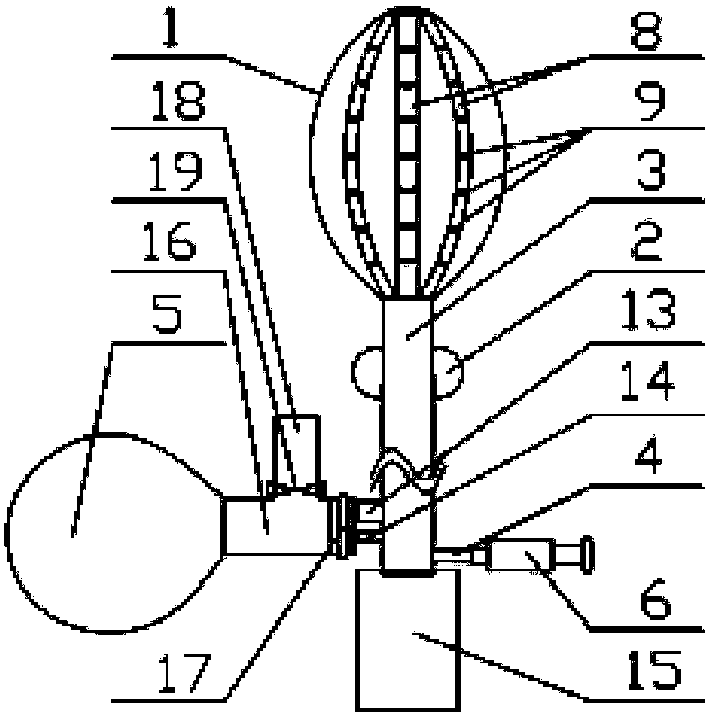
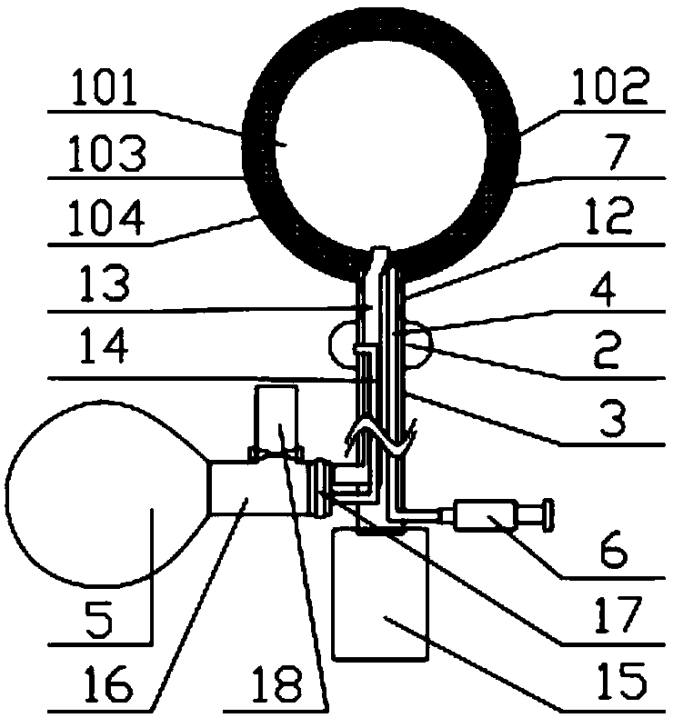
[0031] Such as figure 1 and figure 2 A hemostatic sac for obstetrics and gynecology is shown, including a uterine cavity sac 1, a vaginal sac 2, an inflation tube, a sewage pipe 3, a drug delivery tube 4, an inflatable air bag 5 and a drug syringe 6, and the uterine cavity 1 Comprising an inner capsule body 101 and a drug delivery cavity 102 wrapped on its outer surface, the drug delivery cavity 102 is filled with a cotton material layer 7, the drug syringe 6, the drug delivery tube 4 and the drug delivery cavity 102 are connected in sequence, and the inflatable The air bag 5, the inflation tube and the inner bag body 101 are connected in sequence,

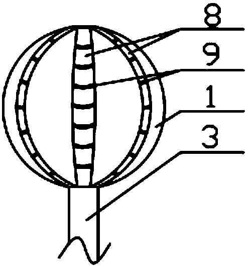
[0032] Such as figure 2 and Figure 5 As shown, the outer wall of the drug administration cavity 102 is a double-layer membrane structure, which are capsule one 103 and capsule two 104 on the outside respectively, as shown in FIG. figure 1 , image 3 and Figure 4 As shown, the outer surface of the capsule 2 104 is equidis...


PUM


Abstract

The invention discloses a hemostatic bag for the obstetrics and gynecology department and belongs to the field of medical instruments. The hemostatic bag comprises a uterine cavity bag body, a vaginalbag body, an inflation tube, a dirt discharging tube, a medicine feeding tube, an inflation airbag and a medicine injector, the outer side wall of a medicine feeding cavity comprises a first bag membrane and a second bag membrane, the second bag membrane is provided with a plurality of diversion grooves connected with the front and rear ends of the uterine cavity bag body, and each diversion groove is internally provided with a series of elastic curved rods whose elastic modulus increases from the front end to the rear end of the uterine cavity bag body; medicine passing micropores are uniformly distributed in the bottoms of the diversion grooves and the outer surface of the second bag membrane respectively, and a plurality of strip-shaped holes parallel to the diversion grooves are uniformly formed in the circumference of the first bag membrane; the rear end of the uterine cavity bag body is connected with the dirt discharging tube which is sleeved with the vaginal bag body, and thedirt discharging tube is provided with a diversion port between the uterine cavity bag body and the vaginal bag body. According to the hemostatic bag, control over discharge of dirt is relatively clear and simple, and closed discharge of the dirt can be achieved completely; the dirt can be prevented from infiltrating into a medicine feeding cavity before inflation, and convenience is provided forensuring a hemostatic function of medicine.

Description

technical field [0001] The invention relates to the field of medical devices, in particular to an obstetrics and gynecology hemostatic bag. Background technique [0002] In clinical obstetrics and gynecology, postpartum hemorrhage is the most common complication, and any hemodynamic instability after delivery is defined as postpartum hemorrhage. At present, the commonly used treatment methods are uterine cavity gauze filling and uterine cavity water bag filling. The process of uterine cavity filling takes a long time, and the pain stimulation to the parturient is severe. It is difficult for patients to bear it under non-anesthesia state, and vaginal packing The procedure cannot avoid gauze contamination, which greatly increases the risk of intrauterine infection. Due to the relaxation of the cervix after delivery, the water sac in the uterine cavity is easy to slip out, resulting in the failure of compression and hemostasis, and the expansion of the water sac is uniform, an...

Claims


Application Information

Patent Timeline
Patent Type & Authority Applications(China)
IPC IPC(8): A61B17/12A61B17/42A61M31/00
CPCA61B17/12A61B17/42A61B17/4241A61B2017/4216A61B2217/005A61M31/00A61M2210/1433A61M2210/005
Inventor 傅珂隋蕾
Owner 武汉半边天医疗技术发展有限公司
Learn More