CT contrast image kidney tumor segmentation method and system based on a three-dimensional convolution neural network

A technology of contrast-enhanced image and neural network, which is applied in the field of medical image processing, can solve the problems of difficult segmentation of kidney tumors and poor segmentation effect, and achieve the effect of enhancing network learning ability, improving segmentation effect, and improving segmentation accuracy

Active Publication Date: 2018-12-18
SOUTHEAST UNIV
View PDF4 Cites 29 Cited by
  • Summary
  • Abstract
  • Description
  • Claims
  • Application Information

AI Technical Summary

Problems solved by technology

[0011] The technical problem to be solved by the present invention: Aiming at the existing problems of difficult segmentation and poor segmentation effect of renal

Method used


Image

  • CT contrast image kidney tumor segmentation method and system based on a three-dimensional convolution neural network
  • CT contrast image kidney tumor segmentation method and system based on a three-dimensional convolution neural network
  • CT contrast image kidney tumor segmentation method and system based on a three-dimensional convolution neural network

Examples


Embodiment

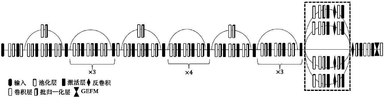
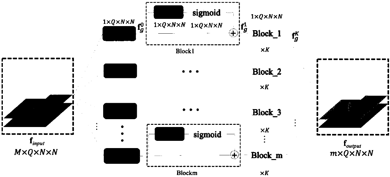
[0069] Embodiment: A three-dimensional deep neural network based on a fully convolutional network proposes to mix continuous two-dimensional CT slices or continuous texture information in MR images. Experimental results show that 3D neural networks generally have better performance than 2D convolutional neural networks in segmentation tasks of different organs, such as liver tumors, brain tumors, lumbar spine, confocal laser microscopy images, etc. After introducing the specific steps and models of the present invention, the test results of the invention on the data set are shown below.

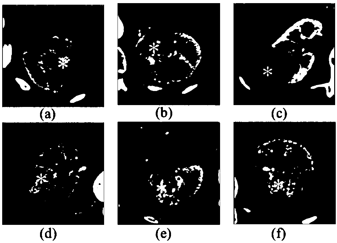
[0070] The experiment uses the CT contrast images obtained in cooperation with the Radiology Department of Jiangsu Provincial People's Hospital. The initial data is 14 patients, and the size is 512×512×200. Because in the CT images of the original patients, irrelevant background areas occupy a large volume, here Some preprocessing was done on the data. Figure 4 It is a 3-dimensional CT cont...


PUM


Abstract

The invention discloses a CT contrast image kidney tumor segmentation method based on a three-dimensional convolution neural network. The method firstly roughly segments the kidney region in the CT contrast image, the kidney and tumor are labeled separately, and the data set is generated. Then the training set is put into the convolution neural network based on pyramid cisternization and step-by-step feature enhancement module, and the training model is obtained. The new kidney data is predicted by the training model, and the segmentation mask of kidney tumor is obtained. The invention also provides a CT contrast image kidney tumor segmentation system based on a three-dimensional convolution neural network. The invention mainly solves the problem of difficult image segmentation of kidney tumor, and the invention can directly obtain a segmentation mask of kidney tumor.

Description

technical field [0001] The invention relates to a medical image processing technology, which belongs to the field of computer application technology. Background technique [0002] Kidney cancer is one of the ten most common cancers in humans. In recent years, traditional radical nephrectomy (RN) is increasingly replacing minimally invasive laparoscopic partial nephrectomy (LPN) for the clinical treatment of local renal cancer. [1] . LPN surgery can remove kidney tumors and preserve normal kidney tissue. In particular, the newly proposed partial resection surgery based on renal artery occlusion technique can preserve renal function to the greatest extent [2] . In order to perform LPN surgery, some useful information, such as tumor size, location, renal anatomy, renal artery and ureter, etc., should be obtained from CT images before surgery. However, manually delineating more than 200 CT slices is a time-consuming and labor-intensive task. Therefore, automatic or semi-au...

Claims


Application Information

Patent Timeline
IPC IPC(8): G06T7/00G06T7/11
CPCG06T7/0012G06T2207/10081G06T2207/20081G06T2207/20084G06T7/11
Inventor 杨冠羽潘覃李国清周忠稳王传霞孔佑勇伍家松杨淳沨舒华忠罗立民
Owner SOUTHEAST UNIV
Why Eureka
Try Eureka