A mobile flat-panel CT imaging system for detecting brain soft tissue
A CT imaging and soft tissue technology, applied in the medical field, can solve the problems of reducing soft tissue contrast, poor image uniformity, and easy misdiagnosis, etc., and achieve stable rotation speed, light weight, and avoid aliasing artifacts
- Summary
- Abstract
- Description
- Claims
- Application Information
AI Technical Summary
Problems solved by technology
Method used
Image
Examples
Embodiment 1
[0038] A method for using a mobile flat-panel CT imaging system for brain soft tissue detection, comprising the following steps:
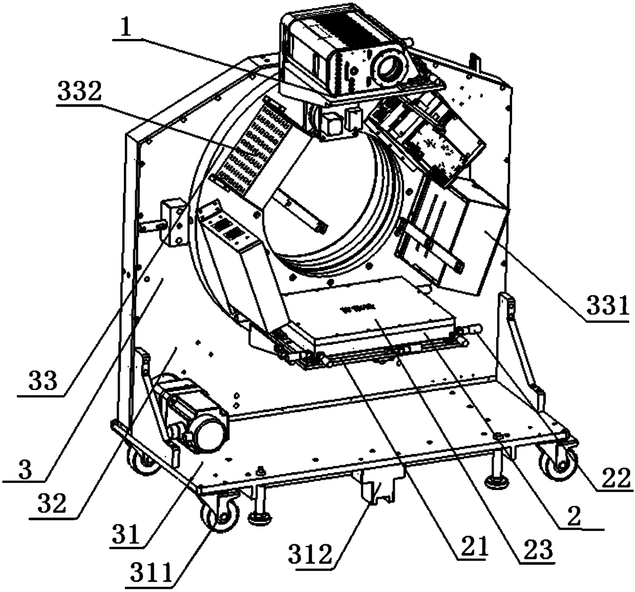
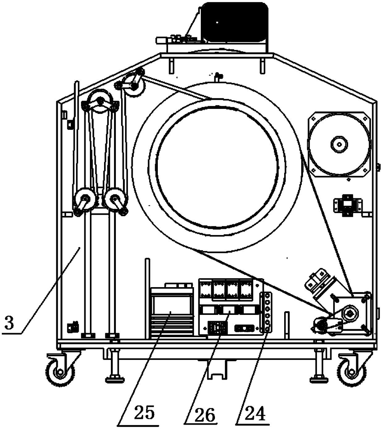
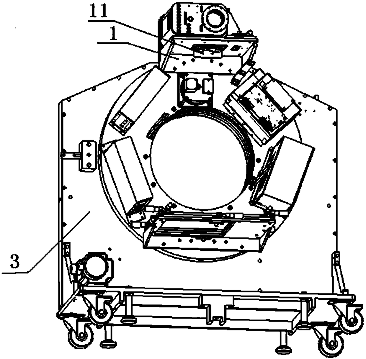
[0039] (1) The flat panel detector 2 is installed on the turntable 33 through the rotating bracket 32, and the redundant radiation shielding layer 26 is arranged behind the flat panel detector 2 to reduce the X-ray dose in the environment, so that Operators will not be exposed to radiation doses; the focal distance between the surface of the flat panel detector 2 and the X-ray source 1 is 600mm, and the installation accuracy is controlled at 0mm. Fine-tuning the X-axis direction and the Z-axis direction of the rotating bracket 32 uses positioning Pin fixed installation position;
[0040] (2) The focal point of the X-ray source 1 is 500 mm from the center of rotation, and the installation accuracy is controlled at 0 mm. The X-ray source 1 is installed on the turntable 33 through the rotating bracket 32 , and the X-ray source 1 passes The X-axis...
Embodiment 2
[0043] A method for using a mobile flat-panel CT imaging system for brain soft tissue detection, comprising the following steps:
[0044] (1) The flat panel detector 2 is installed on the turntable 33 through the rotating bracket 32, and the redundant radiation shielding layer 26 is arranged behind the flat panel detector 2 to reduce the X-ray dose in the environment, so that Operators will not be exposed to radiation doses; the focal distance between the surface of the flat panel detector 2 and the X-ray source 1 is 800mm, and the installation accuracy is controlled at 1mm. Fine-tuning the X-axis direction and the Z-axis direction of the rotating bracket 32 uses positioning Pin fixed installation position;
[0045] (2) The focal point of the X-ray source 1 is 515mm from the center of rotation, and the installation accuracy is controlled at 1mm. The X-ray source 1 is installed on the turntable 33 through the rotating bracket 32, and the X-ray source 1 passes through the The...
Embodiment 3
[0048] A method for using a mobile flat-panel CT imaging system for brain soft tissue detection, comprising the following steps:
[0049] (1) The flat panel detector 2 is installed on the turntable 33 through the rotating bracket 32, and the redundant radiation shielding layer 26 is arranged behind the flat panel detector 2 to reduce the X-ray dose in the environment, so that Operators will not be exposed to radiation doses; the focal distance between the surface of the flat panel detector 2 and the X-ray source 1 is 1000mm, and the installation accuracy is controlled at 2mm. Fine-tuning the X-axis direction and the Z-axis direction of the rotating bracket 32 uses positioning Pin fixed installation position;
[0050] (2) The focal point of the X-ray source 1 is 530mm from the center of rotation, and the installation accuracy is controlled at 2mm. The X-ray source 1 is installed on the turntable 33 through the rotating bracket 32, and the X-ray source 1 passes through the Th...
PUM
| Property | Measurement | Unit |
|---|---|---|
| Thickness | aaaaa | aaaaa |
Abstract
Description
Claims
Application Information
 - R&D Engineer
- R&D Manager
- IP Professional
- Industry Leading Data Capabilities
- Powerful AI technology
- Patent DNA Extraction
Browse by: Latest US Patents, China's latest patents, Technical Efficacy Thesaurus, Application Domain, Technology Topic, Popular Technical Reports.
© 2024 PatSnap. All rights reserved.Legal|Privacy policy|Modern Slavery Act Transparency Statement|Sitemap|About US| Contact US: help@patsnap.com


