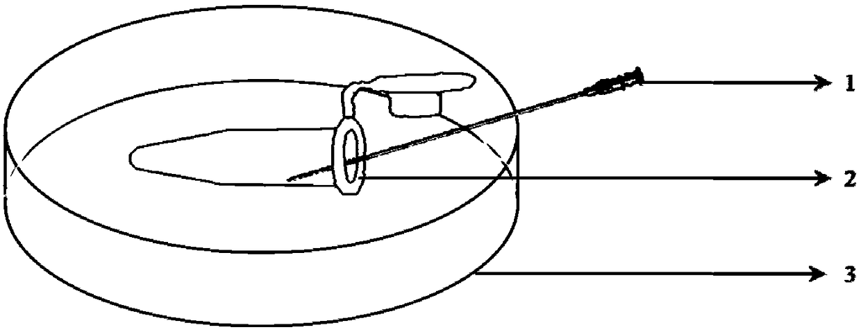
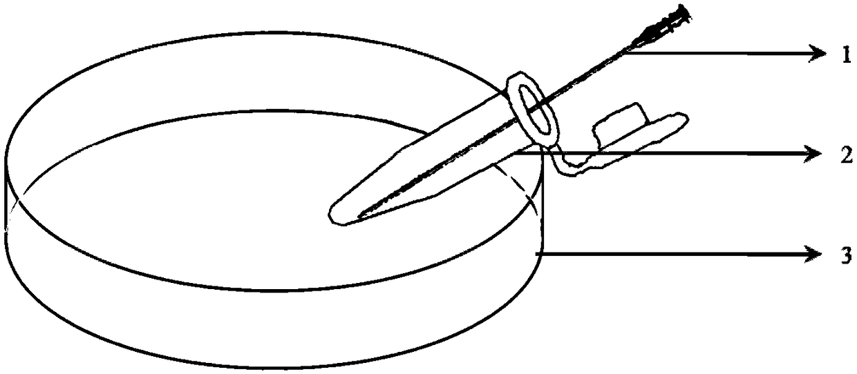
Loading method of biopsy specimens
A biopsy specimen and biopsy technique, applied in the field of biopsy specimens, can solve the problems of specimen loss, easy contamination, and long specimen transfer time, and achieve the effects of saving transfer time, reducing the risk of contamination, and reducing the probability of specimen loss.
- Summary
- Abstract
- Description
- Claims
- Application Information
AI Technical Summary
Problems solved by technology
Method used
Image
Examples
Embodiment 1
[0043] Embodiment 1: a kind of loading method of biopsy specimen,
[0044] S1: Biopsy Dish Preparation
[0045] Prepare at least one Falcon 1006 dish as an embryo biopsy dish, each biopsy dish can biopsy 1 to 5 embryos, place 1 embryo in a single microdroplet, the volume of each microdroplet is 18 μL, cover it with mineral oil and put it into culture box, and mark each droplet with the same number as the number of the biopsy embryo;
[0046] S2: Biopsy dish preparation
[0047] Prepare at least one Falcon 1006 dish as an embryo biopsy washing dish, add 5 washing microdroplets to each biopsy washing dish, and the volume of each microdroplet is 18 μL;
[0048] S3: Blastomere biopsy
[0049] The laser method is used to punch holes in the zona pellucida, and the fixed needle of the micro-operating system is used to fix the embryos, so that the gap in the zona pellucida is located at the 2 o'clock direction, and a flat biopsy needle with an inner diameter of 30um is used to enter ...
Embodiment 2
[0058] Embodiment 2: a kind of loading method of biopsy specimen,
[0059] S1: Biopsy Dish Preparation
[0060] Prepare at least one Falcon 1006 dish as an embryo biopsy dish. Each biopsy dish can biopsy 1 to 5 embryos. Place 1 embryo in a single microdroplet. The volume of each microdroplet is 20 μL. Cover it with mineral oil and put it into culture. box, and mark each droplet with the same number as the number of the biopsy embryo;
[0061] S2: Biopsy dish preparation
[0062] Prepare at least one Falcon 1006 dish as an embryo biopsy washing dish, add 6 washing microdroplets to each biopsy washing dish, and the volume of each microdroplet is 20 μL;
[0063] S3: Blastomere biopsy
[0064] The laser method is used to punch holes in the zona pellucida, and the microscopic operating system is used to fix the embryos, so that the gap in the zona pellucida is located at 2 o'clock, and a flat biopsy needle with an inner diameter of 33um is used to enter from the gap in the zona ...
Embodiment 3
[0073] Embodiment 3: a kind of loading method of biopsy specimen,
[0074] S1: Biopsy Dish Preparation
[0075] Prepare at least one Falcon 1006 dish as an embryo biopsy dish. Each biopsy dish can biopsy 1 to 5 embryos. Place 1 embryo in a single microdroplet. The volume of each microdroplet is 22 μL. Cover with mineral oil and place in culture box, and mark each droplet with the same number as the number of the biopsy embryo;
[0076] S2: Biopsy dish preparation
[0077] Prepare at least one Falcon 1006 dish as an embryo biopsy washing dish, add 7 washing microdroplets to each biopsy washing dish, and the volume of each microdroplet is 22 μL;
[0078] S3: Blastomere biopsy
[0079] The zona pellucida is perforated by the laser method, and the embryo is fixed with a microscopic operating system fixing needle, so that the gap in the zona pellucida is located at the 2 o'clock direction, and a flat biopsy needle with an inner diameter of 35um is used to enter from the gap in t...
PUM
 Abstract
Description
Claims
Application Information
 - R&D
- Intellectual Property
- Life Sciences
- Materials
- Tech Scout
- Unparalleled Data Quality
- Higher Quality Content
- 60% Fewer Hallucinations
Browse by: Latest US Patents, China's latest patents, Technical Efficacy Thesaurus, Application Domain, Technology Topic, Popular Technical Reports.
© 2025 PatSnap. All rights reserved.Legal|Privacy policy|Modern Slavery Act Transparency Statement|Sitemap|About US| Contact US: help@patsnap.com


