A Retinal Vessel Segmentation Method Based on Convolutional Neural Network

A convolutional neural network and retinal blood vessel technology, applied in image analysis, image enhancement, instrumentation, etc., can solve problems that are not suitable for practical applications, complex preprocessing and postprocessing steps, and avoid training overfitting and amplification The method is simple and the effect of improving accuracy

Active Publication Date: 2021-08-10
SOUTH CHINA UNIV OF TECH
View PDF0 Cites 0 Cited by
  • Summary
  • Abstract
  • Description
  • Claims
  • Application Information

AI Technical Summary

Problems solved by technology

[0006] Many of the above methods for retinal vessel segmentation, some are not suitable for practical applications, some require more complex preprocessing and postprocessing steps, and want to be applied to computer-aided diagnosis, the accuracy of the algorithm to achieve retinal vessel segmentation, Sensitivity, specificity, etc. have certain requirements

Method used


Image

  • A Retinal Vessel Segmentation Method Based on Convolutional Neural Network
  • A Retinal Vessel Segmentation Method Based on Convolutional Neural Network
  • A Retinal Vessel Segmentation Method Based on Convolutional Neural Network

Examples


Embodiment

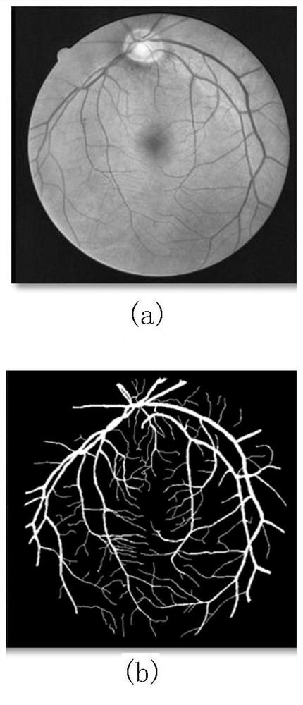
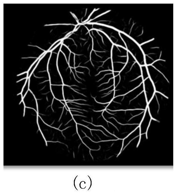
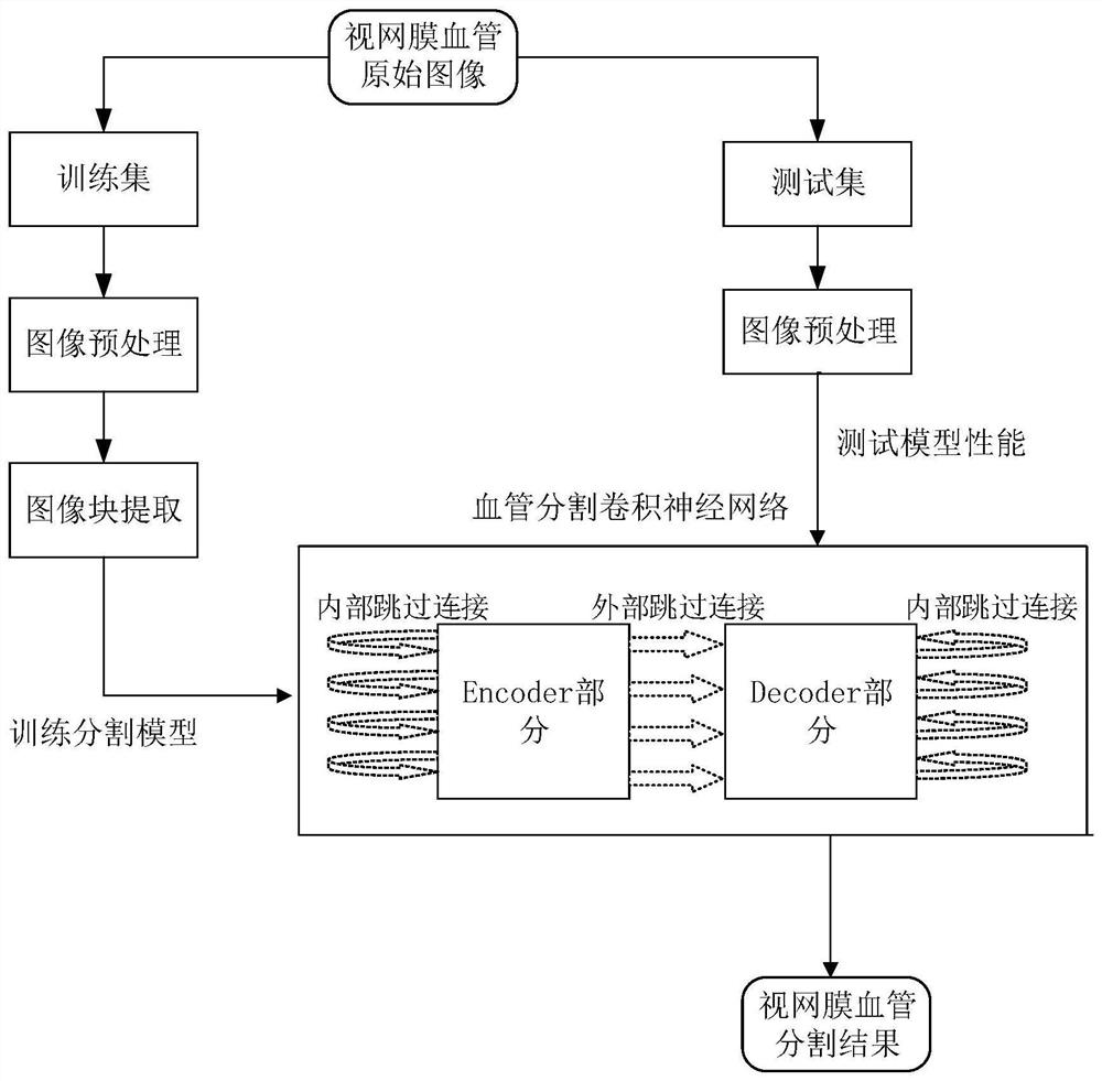
[0035] The overall segmentation flow chart of this embodiment is as follows figure 1 shown. In this embodiment, the DRIVE (Digital Retinal Image for Vessel Extraction) public database is used as the experimental data. There are 40 retinal fundus images in the database, which are divided into a training set and a test set, each of which has 20 images. In the training set, each retinal image has an original image and a corresponding expert manual segmentation map (groundtruth), and the expert segmentation result is used as the standard, that is, the label of the training data, for the training of the network model. Each original retinal image in the test set has manual segmentation maps corresponding to two experts. During the test, the segmentation result of the first expert is used as the true value to evaluate the segmentation performance of the model proposed by the present invention. Various obtained The index value is compared with the segmentation result of the second ex...


PUM


Abstract

The invention discloses a retinal blood vessel segmentation method based on a convolutional neural network, which includes: preprocessing the retinal fundus image; performing block extraction on training set images; constructing a convolutional neural network for blood vessel segmentation, and using the extracted image blocks to perform Training; in the prediction stage, multiple consecutive overlapping segments are extracted for each image, and the classification probability of each pixel is obtained by averaging multiple prediction results, and the final segmentation result map is obtained. The new convolutional neural network structure proposed by the present invention for retinal vessel segmentation is a symmetrical network based on the Encoder-Decoder structure, and two skip connections are added between the Encoder part and the Decoder part. The network can not only realize the end-to-end segmentation of retinal images, but also can obtain accurate segmentation results on a limited data set, and can effectively avoid the problem of gradient disappearance, which has certain advantages compared with existing algorithms.

Description

technical field [0001] The invention relates to the technical fields of medical image processing and computer vision, in particular to a retinal vessel segmentation method based on a convolutional neural network. Background technique [0002] Retinal fundus images have been widely used in the diagnosis, screening and treatment of various cardiovascular and ophthalmic diseases, and the analysis of retinal blood vessels is of great significance in revealing important information of systemic diseases in many clinical applications. Segmentation of retinal vessels is a fundamental step in quantitative analysis. The segmented vascular tree can be used to extract morphological attributes of vessels, such as length, width, branching and angle. Furthermore, the vascular tree, as the most stable feature in an image, has been adopted in multimodal retinal image registration, and due to its uniqueness, the vascular tree has also been used in biometrics. Manual segmentation of vascular...

Claims


Application Information

Patent Timeline
Patent Type & Authority Patents(China)
IPC IPC(8): G06T7/10
CPCG06T2207/20081G06T2207/20084G06T2207/30041G06T2207/30101G06T7/10
Inventor 周叶萍陆以勤覃健诚
Owner SOUTH CHINA UNIV OF TECH
Why Eureka
Try Eureka