DNA quantitative analysis method based on microscope images of cells

A quantitative analysis and microscopy technology, applied in the field of image processing, can solve the problems of affecting accuracy, time-consuming, missing diploid cells, etc., and achieve the effect of improving accuracy and stabilizing results

Inactive Publication Date: 2019-03-29
湖南品胜生物技术有限公司
View PDF10 Cites 5 Cited by
  • Summary
  • Abstract
  • Description
  • Claims
  • Application Information

AI Technical Summary

Problems solved by technology

However, this type of method will miss a large number of diploid cells when dealing with densely distributed nuclei and large changes in the gray distribution of the nuclei, and often extracts all the features of each candidate area when identifying them. time-consuming in the case of
[0004] The source steps of the picture in the current DNA quantitative analysis method are as follows: incident light → glass slidetransmitted light → CCD (or CMOS), and finally the CCD (or CMOS) restores the color of the picture to form a color picture or a black picture , when calculating the IOD value in DNA, it is necessary to compensate the RGB value of the picture during the color restoration process of the picture or when calculating the gray value of the picture after grayscale, so accurate IOD cannot be obtained value, so that it will affect the accuracy of DNA quantitative analysis

Method used


Image

  • DNA quantitative analysis method based on microscope images of cells

Examples


Embodiment Construction

[0018] The present invention will be described in detail below with reference to the drawings in the embodiments of the present invention.

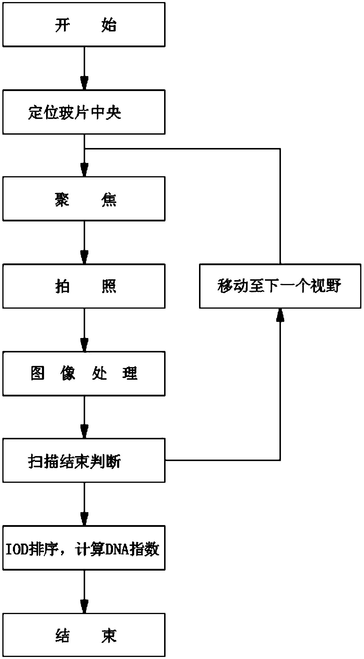
[0019] Such as figure 1 As shown, a DNA quantitative analysis method based on a cell microscope image disclosed in the present invention comprises the following steps:

[0020] Step 1: Position the cell-laden slide in the detection center of the microscope stage so that the cell-enriched area is under the observation line of sight of the microscope;

[0021] Step 2: Focus on the cell plane and take a picture, save the picture after taking the picture, and use the information file directly obtained from the CCD or CMOS as the analysis picture;

[0022] Further, the information files obtained directly from the CCD or CMOS are RAW files.

[0023] Step 3: Analyze the image after taking the photo: collect normal diploid, abnormal ploid, lymphocyte, and other cell images, and calculate the integral optical density value of the nucleus after i...


PUM


Abstract

The invention discloses a DNA quantitative analysis method based on microscope images of cells. According to the DNA quantitative analysis method based on the microscope images of cells, an information RAW file obtained directly from a CCD or a CMOS is used as an image for the DNA quantitative analysis, so as to avoid the errors resulting from the increased compensation calculations when calculating an IOD value, to greatly improve the accuracy of DNA quantitative calculation, and to make the calculation result to be more stable when the intensity of the incident light is increased or decreased.

Description

technical field [0001] The invention relates to the technical field of image processing, in particular to a DNA quantitative analysis method based on cell microscope images. Background technique [0002] Cytological examination is the main way for early screening of typical cancers such as cervical cancer and oral cancer. The smear technique used in the early days was popular because of its low price, but its missed diagnosis rate is high. Although the liquid-based smear technique widely used since the 21st century The effect of film production has been improved, but it requires experienced pathologists to directly observe the morphology of cells under a microscope to give diagnostic results, which is inefficient, easily affected by subjectivity, and has poor repeatability. DNA ploidy analysis technology automatically analyzes the nucleus in the cell image, and then quantitatively measures the chromosome or DNA content in the nucleus, and automatically gives the judgment res...

Claims


Application Information

Patent Timeline
IPC IPC(8): G01N21/84
CPCG01N21/84
Inventor 贺权源刘朝前李仕能
Owner 湖南品胜生物技术有限公司
Why Eureka
Try Eureka