A method for cryopreservation and recovery of three-dimensional retinal tissue

A three-dimensional retina and cryopreservation technology, applied in the field of cryopreservation and recovery of three-dimensional retinal tissue, can solve the problems of long recovery time, low success rate, nerve axon rupture, etc., and achieve high success rate and good repeatability Effect

Active Publication Date: 2022-07-26
ZHONGSHAN OPHTHALMIC CENT SUN YAT SEN UNIV
View PDF0 Cites 0 Cited by
  • Summary
  • Abstract
  • Description
  • Claims
  • Application Information

AI Technical Summary

Problems solved by technology

However, these two cryopreservation methods have problems such as poor reproducibility, low success rate, long recovery time after resuscitation, disordered retinal tissue structure after resuscitation, and nerve axon rupture.
[0005] At present, there is still no satisfactory cryopreservation and recovery method for three-dimensional retinal tissue. The existing reports all have the problem of failure to recover successfully or poor recovery of tissue structure and function after recovery, which seriously restricts the application of three-dimensional retinal tissue in drug screening and gene manipulation. and cell replacement therapy

Method used


Image

  • A method for cryopreservation and recovery of three-dimensional retinal tissue
  • A method for cryopreservation and recovery of three-dimensional retinal tissue
  • A method for cryopreservation and recovery of three-dimensional retinal tissue

Examples


Embodiment 1 3

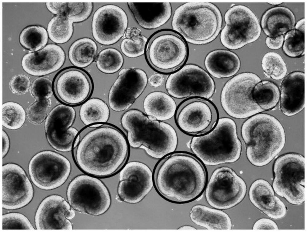
[0035] Example 1 Cryopreservation of three-dimensional retinal tissue

[0036](1) Cells: 3D retinal tissue obtained by differentiation of BC1-GFP (induced pluripotent stem cells derived from blood) (the 6th week of differentiation). For the specific preparation method, please refer to Zhong X, Gutierrez C, Xue T, Hampton C, VergaraMN , Cao LH, et al.Generation of three-dimensional retinal tissue with functional photoreceptors from human iPSCs.Nature communications.2014;5:4047.

[0037] (2) Reagents and consumables:

[0038] ①RDM medium: DMEM / F12 (C11330500BT, Gibco, 4℃), DMEM (Gibco, C11995500BT, 4℃), B27 (12587010, Gibco, -20℃), NEAA (11140-050, Gibco, 4℃), Antibiotic -antimycotic(15240,Gibco,-20℃);

[0039] ②DMSO: D4540, Sigma-Aldrich, room temperature;

[0040] ③Sucrose: S0389, Sigma-Aldrich, room temperature;

[0041] ④Blebbitatin: B0560, Sigma-Aldrich, -20℃;

[0042] ⑤FBS (calf serum): 10099-141, Gibco, -20℃;

[0043] ⑥Small dish: 430165, Corning;

[0044] ⑦Cryopre...

Embodiment 2 3

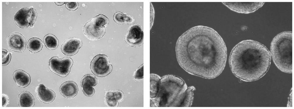
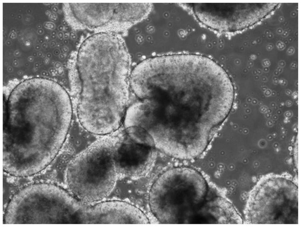
[0056] Example 2 Resuscitation of three-dimensional retinal tissue

[0057] (1) The composition and preparation of the recovery medium:

[0058] The composition of the recovery medium: DMEM mixed medium supplemented with 2% (v / v) B27, 1% (v / v) NEAA, 1% (v / v) Antibiotic-Antimycotic and 20% (v / v) small Bovine serum, DMEM mixed medium is DMEM / F12:DMEM=3:2 (v / v); it is formulated as: DMEM / F12 and DMEM are mixed in a volume ratio of 3:2 to obtain DMEM mixed medium, and then mixed with DMEM Add B27, NEAA, Antibiotic-Antimycotic, calf serum to the medium, mix well, apply filter sterilization, and store in a refrigerator at 4°C or -20°C. Store for 6 months and rewarm in a 37°C water bath before use.

[0059] (2) Recovery steps:

[0060] Take out the cryopreservation tube (containing 3D retinal tissue) saved in Example 1 from liquid nitrogen, quickly add 1.3 mL of 37°C resuscitation culture medium, place it in a 37°C water bath to quickly thaw and shake to mix the liquid; carefully ...


PUM


Abstract

The invention discloses a cryopreservation and recovery method of three-dimensional retinal tissue. The cryopreservation steps of the method are as follows: select the 3D retinal tissue suitable for cryopreservation, suspend it in RDM medium, then transfer it to a cryopreservation tube, add 2× cryopreservation solution in an equal volume ratio, and wait for the 3D retinal tissue to sink to the bottom of the tube Aspirate part of the supernatant, seal the cryopreservation tube, place it at -80°C overnight to cool down, and then transfer it to liquid nitrogen for storage; finally, the preserved retinal tissue can be thawed and thawed and placed in an incubator for suspension culture. The method provided by the invention has good repeatability and high success rate, and the resuscitation survival rate can be as high as 80-90%.

Description

technical field [0001] The invention relates to the technical field of stem cell induced differentiation and tissue cryopreservation, in particular to a method for cryopreservation and recovery of three-dimensional retinal tissue obtained by inductive differentiation of human pluripotent stem cells. Background technique [0002] At present, breakthroughs in stem cell regeneration technology have brought unprecedented opportunities for research on the pathogenesis of refractory diseases, new drug development, and cell therapy. Among them, breakthroughs have been made in the key technology of inducing human pluripotent stem cells (hPSCs) to differentiate into retinal tissues with three-dimensional structure in vitro, becoming the leader of in vitro regeneration of stem cells, and relevant clinical experiments are also in full swing. [0003] The regenerated retinal tissue is an organoid with a compact, three-dimensional structure and is developmentally part of the nervous syst...

Claims


Application Information

Patent Timeline
Patent Type & Authority Patents(China)
IPC IPC(8): C12N5/071A01N1/02
CPCA01N1/0221A01N1/0226C12N5/0621
Inventor 葛坚罗子明冼碧琨李凯婧叶美芳
Owner ZHONGSHAN OPHTHALMIC CENT SUN YAT SEN UNIV
Why Eureka
Try Eureka