One-step fluorescence detection system and thrombin detection method

A technology for fluorescence detection and thrombin, applied in the field of analytical chemistry, can solve the problem of low sensitivity

Active Publication Date: 2019-06-14
SICHUAN UNIV
View PDF2 Cites 2 Cited by
  • Summary
  • Abstract
  • Description
  • Claims
  • Application Information

AI Technical Summary

Problems solved by technology

However, these detection methods need to label probes and prepare nanoparticles, and the sensitivity is also low

Method used


Image

  • One-step fluorescence detection system and thrombin detection method
  • One-step fluorescence detection system and thrombin detection method
  • One-step fluorescence detection system and thrombin detection method

Examples


preparation example Construction

[0032]In the detection system of the present invention, the preparation method of the B-H2 functionalized nanofiber membrane comprises the following steps: (1) dissolving in N, N-dimethyl ammonium bromide after mixing polystyrene and tetrabutylammonium bromide In methyl formamide, obtain PS / TBAB / DMF solution; The mixture quality of described polystyrene and tetrabutylammonium bromide is 15~25% of described PS / TBAB / DMF solution quality;

[0033] (2) The PS / TBAB / DMF solution is subjected to electrospinning, and the PS nanofiber membrane is obtained after drying; the voltage during the electrospinning is 10-20kV, and the injection speed is 2-8 μL / min, and the receiving The distance is 7~15cm, and the collection time is 1.5~3h;

[0034] (3) Utilize the argon gas plasma that dielectric barrier discharge produces to process described PS nanofiber membrane, obtain the PS nanofiber membrane after processing; The voltage of described treatment is 40~50V, and electric current is 1.2~2.5...

Embodiment 1

[0046] The PS nanofiber membrane is prepared by electrospinning technology, and the specific operation is as follows: using DMF as a solvent, prepare a PS / TBAB / DMF solution with a mass fraction of 20%, wherein TBAB is 0.3% (w / v), and stir at room temperature for 24h , after mixing evenly, electrospinning was carried out. Spinning conditions: voltage 15kV, sample injection speed 5μL / min, receiving distance 10cm, collection time 2h, ambient humidity ~49%. The nanofiber membrane received on the aluminum foil was placed in an oven at 80° C. for 4 hours, and then the nanofiber membrane was cut into discs with a diameter of d=5 mm for future use.

[0047] The PS nanofiber membrane was treated by argon plasma generated by dielectric barrier discharge, the voltage was set to 45V, the current was 2.0A, and the discharge treatment time was 2min. Immediately soak the treated PS nanofiber membrane in 100 μL 2.0 μM avidin solution for 1 h at room temperature, take it out and soak it in ul...

Embodiment 2

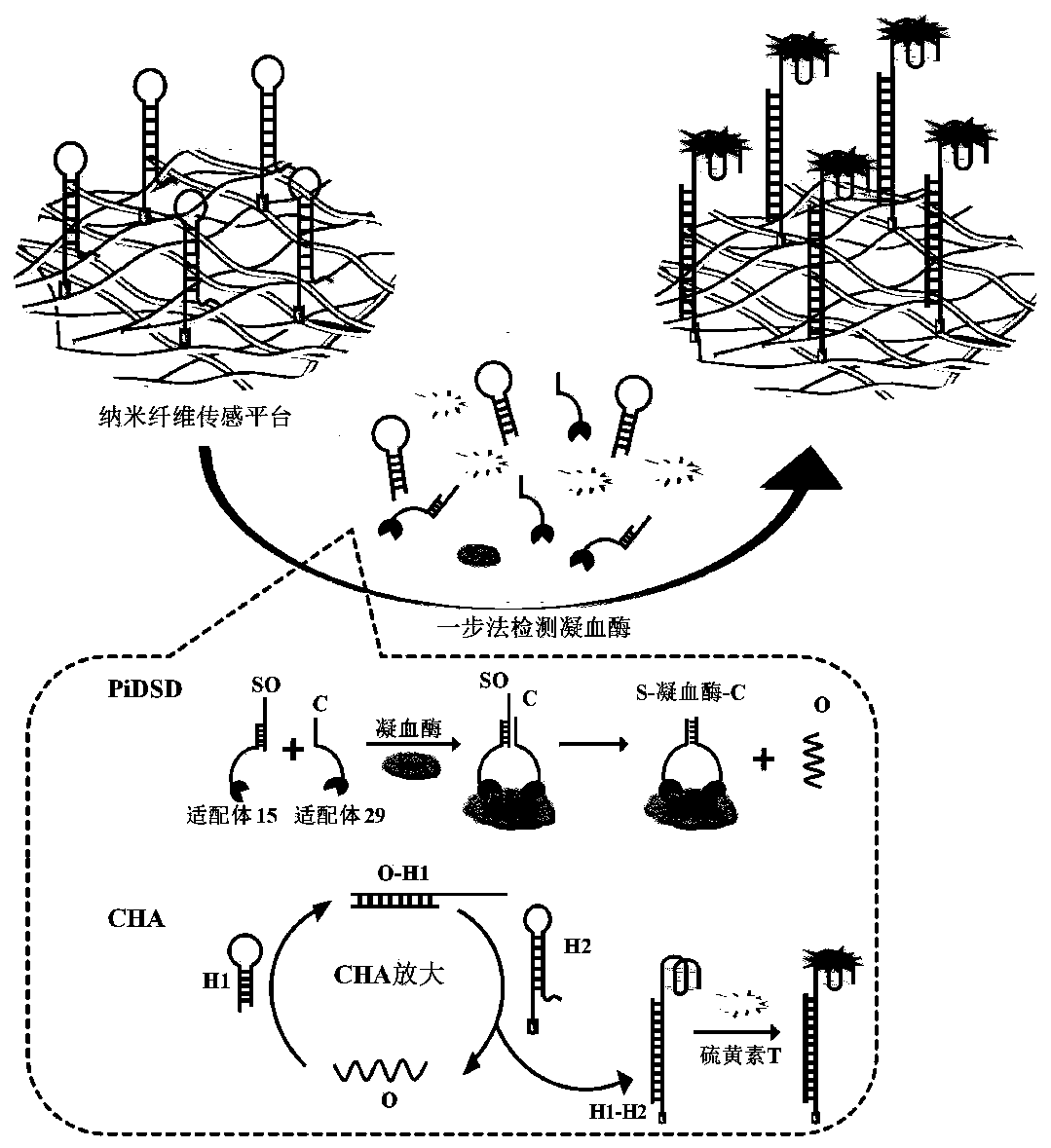
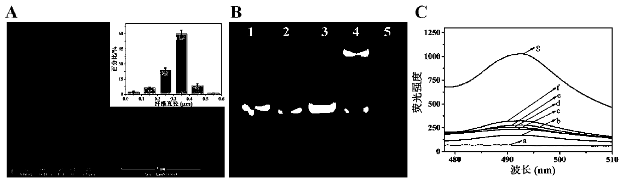
[0049] Proximity-induced DNA strand displacement (PiDSD) process demonstrated by polyacrylamide gel electrophoresis (PAGE)

[0050] Prepare 12% polyacrylamide gel, then mix 8 μL DNA sample with 2 μL 6× gel loading buffer (0.25% bromophenol blue, 0.25% xylene cyanol, 40% (w / v) sucrose solution), Add it to the well of the gel for electrophoresis. Electrophoresis was performed in 1×TBE buffer (89mM Tris, 89mM boric acid, 2mM EDTA, pH=8.3) and a constant voltage of 110V for 1h. At this time, the gel was stained with EB for 30 minutes, and then photographed with a UV imaging system (Clinx GenoSens, China).

[0051] PiDSD results such as figure 2As shown in B, bands 1, 2, and 5 represent SO, C, and O chains, respectively. In the absence of thrombin, the reaction between SO and C chain for 100min could not produce export chain O (band 3). When thrombin is present, a band exporting O chains can form at the bottom of band 4. In addition, a new slow-moving S-thrombin-C band was ge...


PUM

PropertyMeasurementUnit
Wavelengthaaaaaaaaaa

Abstract

The invention provides a one-step fluorescence detection system and a thrombin detection method, and relates to the technical field of analytical chemistry. The detection system comprises a B-H2 functionalized nano-fiber membrane, support DNA, output DNA, competitive DNA, a buffer solution, thioflavin T (ThT), an H1 sequence and thrombin. When the detection system is used for detecting the thrombin, based on the improvement of reaction thermodynamics and kinetics on a surface interface of the nano-fiber membrane, the corresponding detection system has a detection limit of 1.0pM, and has a goodlinear relation between 50pM and 5nM, excellent selectivity and long-term stability. When the detection system is used for detecting a sample, a PiDSD process, CHA amplification and a ThT binding reaction are integrated, so that the separation and elution processes required by ELISA are avoided, the time is shortened, and the steps are simplified.

Description

technical field [0001] The invention belongs to the technical field of analytical chemistry, and in particular relates to a one-step fluorescent detection system and a thrombin detection method. Background technique [0002] Sensitive, convenient, reliable and low-cost detection of protein markers is becoming more and more important in point-of-care testing (POCT) and early diagnosis. Common detection methods such as enzyme-linked immunosorbent assay (ELISA), electrochemical assay, optical method and so on. These methods contain multi-step separation and elution steps, are complicated to operate, require sophisticated instruments, and have characteristics such as relatively low sensitivity. [0003] Electrospun nanofibrous membrane has the advantages of controllable diameter, porous structure, high specific surface area and easy modification, and is considered to be an ideal sensing interface material. Photochemical sensors based on electrospun nanofiber membranes have bee...

Claims


Application Information

Patent Timeline
IPC IPC(8): G01N33/573
Inventor 周翠松李晓玲肖丹
Owner SICHUAN UNIV
Why Eureka
Try Eureka