
Medical image analysis method, device, computer equipment and storage medium

A technology of medical imaging and analysis methods, applied in medical images, image analysis, computer components, etc.

Active Publication Date: 2019-06-21
TENCENT TECH (SHENZHEN) CO LTD
View PDF6 Cites 9 Cited by
  • Summary
  • Abstract
  • Description
  • Claims
  • Application Information

AI Technical Summary

Problems solved by technology

Since it is difficult for a single tissue image to fully reflect the information of the patient's tissue slices, the accuracy of the diagnostic classification results needs to be further improved

Method used


Image

  • Medical image analysis method, device, computer equipment and storage medium
  • Medical image analysis method, device, computer equipment and storage medium
  • Medical image analysis method, device, computer equipment and storage medium

Examples


Embodiment Construction

[0034] In order to make the purpose, technical solution and advantages of the present application clearer, the present application will be further described in detail below in conjunction with the accompanying drawings and embodiments. It should be understood that the specific embodiments described here are only used to explain the present application, and are not intended to limit the present application.

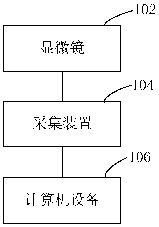
[0035] figure 1 It is a schematic diagram of the application environment of the medical image analysis method in one embodiment. The doctor puts the pathological slice under the microscope 102 to observe the pathological slice. When the doctor observes the slice, the video data under the field of view of the microscope 102 is collected by the collection device 104 to obtain a medical image video stream. The medical image analysis method can be applied in the computer device 106 . The computer device 106 acquires a medical image video stream, and the medical image video ...


PUM


Abstract

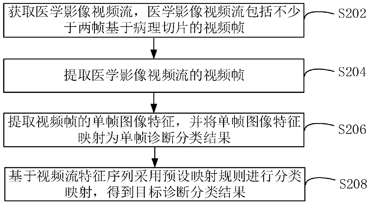
The invention relates to a medical image analysis method, a device, computer equipment and a storage medium. The medical image analysis method comprises the steps of acquiring a medical image video stream which comprises no less than two video frames based on pathological slices; extracting the video frame of the medical image video stream; extracting the single-frame image characteristic of the video frame, mapping the single-frame image characteristic to a single-frame diagnosis classification result; based on a video stream characteristic sequence, performing classified mapping according toa preset mapping rule, and obtaining a target diagnosis classification result; wherein the video stream characteristic sequence comprises the single-frame image characteristic and the single-frame diagnosis classification result of each video frame. Because characteristic extraction and classification mapping are performed based on the medical image video stream for obtaining the target diagnosisclassification result and the medical image video stream comprises no less than two video frames based on the pathological slices, accuracy of the diagnosis classification result can be improved, thereby supplying an accurate basis for medical diagnosis.

Description

technical field [0001] The present application relates to the field of medical image processing, in particular to a medical image analysis method, device, computer equipment and storage medium. Background technique [0002] Medical imaging refers to internal tissue images of the human body or a part of the human body obtained in a non-invasive manner for medical treatment or medical research. The analysis of medical images can provide auxiliary basis for doctors to obtain accurate diagnosis results. [0003] The traditional medical image analysis method analyzes the single tissue image determined by the doctor in the process of observing the pathological section, and obtains the diagnostic classification result corresponding to the tissue image. Since it is difficult for a single tissue image to fully reflect the information of the patient's tissue slices, the accuracy of the diagnostic classification results needs to be further improved. Contents of the invention [000...

Claims


Application Information

Patent Timeline
IPC IPC(8): G16H30/00G16H50/20G06K9/62
CPCG16H50/20G16H30/00G06F18/24G06T7/0012G06T2207/10016G06T2207/10056G06T2207/20081G06T2207/20084G06T2207/30096G02B21/365G06N3/08G06V20/69G06V20/46G06N3/045G06V20/41
Inventor 陆唯佳姚建华韩骁周昵昀
Owner TENCENT TECH (SHENZHEN) CO LTD
Learn More