Surgical tumor operation real-time navigation method based on indocyanogen-green fluorescence development

A navigation method, the technology of indocyanine green, applied in the field of medical image processing, can solve the problems of inability to obtain boundaries, ineffective acquisition of boundaries, lack of scientific quantification, etc.

Inactive Publication Date: 2019-06-25
安徽紫薇帝星数字科技有限公司
View PDF8 Cites 5 Cited by
  • Summary
  • Abstract
  • Description
  • Claims
  • Application Information

AI Technical Summary

Problems solved by technology

At present, ICG has been widely used in endoscopic surgery, but the existing surgical navigation system guided by ICG only simply displays the fluorescence image, and then the doctor observes the image and draws the boundary based on his own clinical experience, which lacks scientific quantification standard
ICG staining is a sequential process, that is, with th

Method used


Image

  • Surgical tumor operation real-time navigation method based on indocyanogen-green fluorescence development
  • Surgical tumor operation real-time navigation method based on indocyanogen-green fluorescence development
  • Surgical tumor operation real-time navigation method based on indocyanogen-green fluorescence development

Examples


Embodiment 1

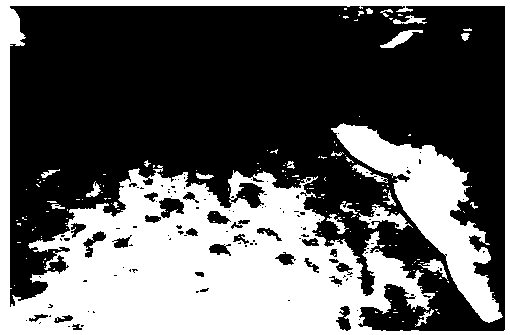
[0019] A real-time navigation method for surgical tumor surgery based on indocyanine green fluorescence imaging, such as figure 1 , including the following steps: 1. Obtain each frame image in the real-time ICG fluorescence surgery video and save it as an ICG fluorescence image; 2. Use the tumor boundary segmentation model based on deep learning to segment the tumor boundary in the ICG fluorescence image, and use Colored lines are added to the original ICG fluorescence image for display, such as figure 2 3. Use the edge evaluation function to evaluate the segmentation boundary of the ICG fluorescence image, and obtain its corresponding boundary clarity coefficient; 4. Find the image with the largest boundary clarity coefficient, and then superimpose its corresponding segmentation boundary under conventional illumination on the tumor image, and display the superimposed results on the screen, such as image 3 As shown, guide the doctor to perform tumor resection.

[0020] The...


PUM


Abstract

The invention discloses a surgical tumor operation real-time navigation method based on indocyanogen-green (Indocyanine-green for English abbreviation (ICG)) fluorescence development. The surgical tumor operation real-time navigation method comprises the following steps: 1, obtaining each frame of image in a real-time ICG fluorescence operation video and storing the image as an ICG fluorescence image; 2, segmenting a tumor boundary in the ICG fluorescence image by using a tumor boundary segmentation model based on deep learning, and adding the tumor boundary to the original ICG fluorescence image in a color line manner for display; 3, evaluating the segmentation boundary of the ICG fluorescence image by adopting an edge evaluation function to obtain a corresponding boundary clearness coefficient; and 4, finding out the image with the maximum boundary clearness coefficient, superposing the corresponding segmentation boundary on the tumor image under the conventional illumination, displaying a superposing result on a screen, and guiding a doctor to carry out a tumor excision operation.

Description

technical field [0001] The invention relates to the technical field of medical image processing, in particular to a real-time navigation method for tumor surgery based on indocyanine green fluorescence imaging. Background technique [0002] China is the largest country with liver cancer, and a large number of patients die of liver cancer every year. Surgical removal of liver cancer cells completely is the most effective way to treat liver cancer. During the operation, surgeons usually use preoperative CT, MRI and other imaging data, and use visual diagnosis, palpation, and intraoperative ultrasound to judge the tumor margin and determine the extent of resection. Due to the problems of huge equipment, ray radiation, and long examination time in CT and MRI, intraoperative real-time imaging evaluation is limited to ultrasonography. However, ultrasonography requires high professional skills in image interpretation and is difficult to popularize in liver surgery. [0003] Indoc...

Claims


Application Information

Patent Timeline
IPC IPC(8): G06T7/11G06T7/12G06T7/187G07C9/00A61B34/20G06N3/08
Inventor 刘荣王斐王子政王宜主张勇王翊
Owner 安徽紫薇帝星数字科技有限公司
Why Eureka
Try Eureka