Medical image fusion method based on improved pulse coupling neural network

A technology of pulse-coupling nerves and medical images, applied in the field of medical image fusion, which can solve the problems of not being able to meet medical needs, not being able to provide complete information on organs or tissue parts at the same time, and subjectively affecting the accuracy.

Active Publication Date: 2019-06-25
JILIN UNIV
View PDF4 Cites 14 Cited by
  • Summary
  • Abstract
  • Description
  • Claims
  • Application Information

AI Technical Summary

Problems solved by technology

If a variety of images only rely on the doctor's spatial conception and speculation to determine the required information, the accuracy will be affected subjectively, and some information may also be ignored
A single medical imaging system can only provide limited information, and cannot provide complete information of an organ or tissue part at multiple angles (or modalities) at the same time, so

Method used


Image

  • Medical image fusion method based on improved pulse coupling neural network
  • Medical image fusion method based on improved pulse coupling neural network
  • Medical image fusion method based on improved pulse coupling neural network

Examples


Embodiment 1

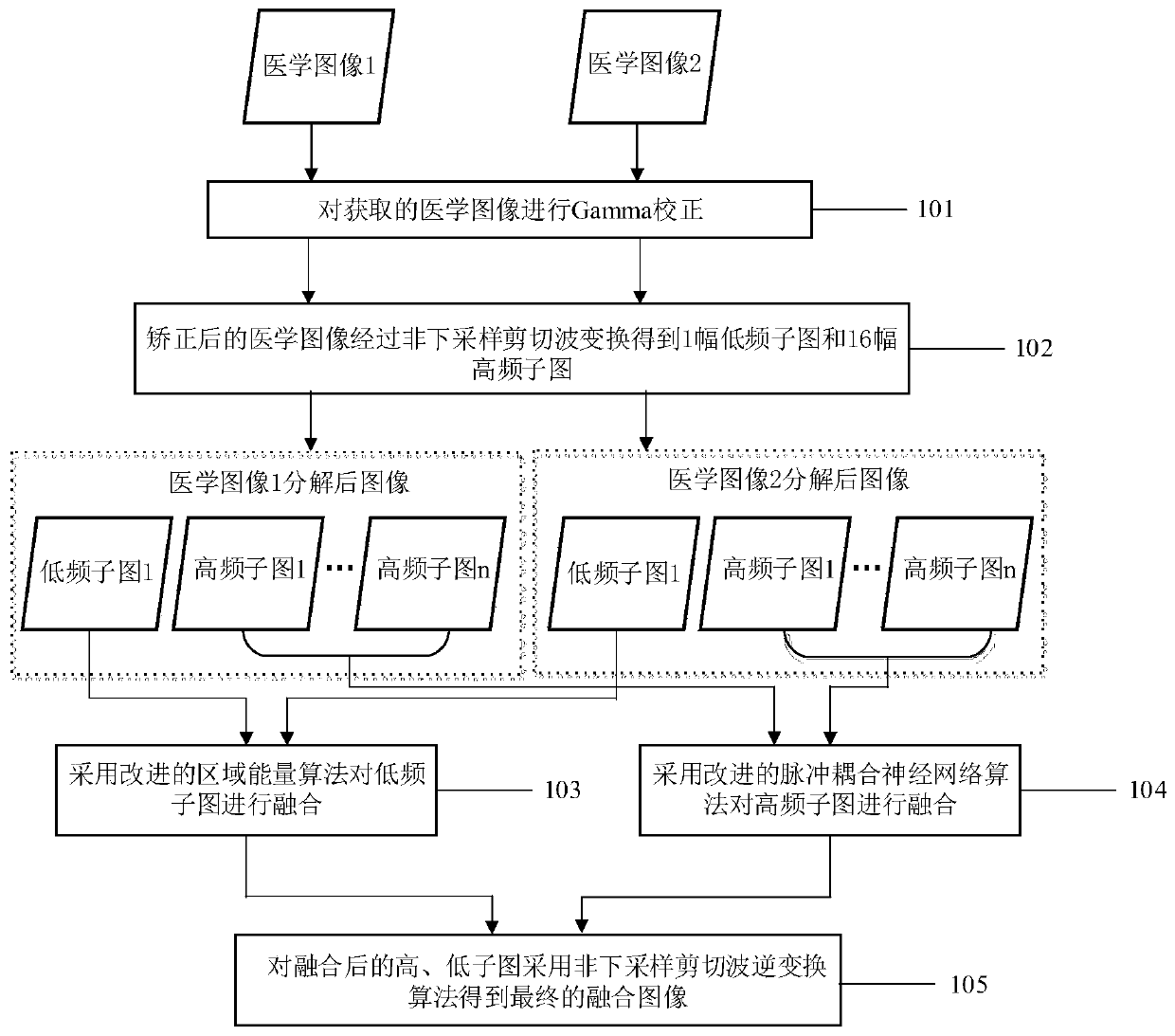
[0150] According to the technical scheme of the present invention, two fully registered medical images are fused. This method is compared with other methods, including image fusion based on multi-scale transform method (NSCT), fusion method based on sparse representation and multi-scale transform (DTCWT-SR), fusion method based on pulse-coupled neural network and multi-scale transform (NSCT-PCNN, NSCT-SF-PCNN), dense SIFT-based image fusion (DSIFT) and boundary-finding-based image fusion (BF). All parameter settings of comparative experiments are default values.

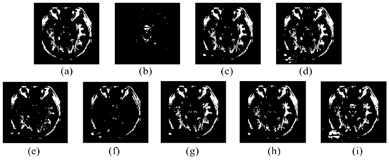
[0151] figure 2 (a) is the nuclear magnetic resonance T1 image (MR-T1), figure 2 (b) is an MRI T2 image (MR-T2), figure 2 (c) is the fusion result graph of the BF method, figure 2 (d) is the fusion result graph of the DSIFT method, figure 2 (e) is the fusion result diagram of the DTCWT-SR method, figure 2 (f) is the fusion result map of the NSCT method, figure 2 (g) is the fusion result map of the NSCT-...

Embodiment 2

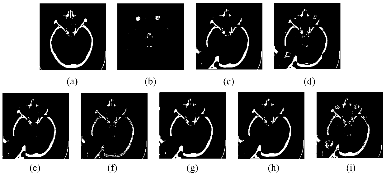
[0157] image 3 Shows a patient with cerebrovascular disease who has been attacked or stroked in the head. The patient only had writing function and lost reading function. The black box indicates the location of the eyeball in the brain, and the lower left corner of the image is a zoomed-in rendering. image 3 (a) is a CT image, image 3 (b) MR image, image 3 (c) is the fusion result graph of the BF method, image 3 (d) is the fusion result graph of the DSIFT method, image 3 (e) is the fusion result diagram of the DTCWT-SR method, image 3 (f) is the fusion result map of the NSCT method, image 3 (g) is the fusion result map of the NSCT-PCNN method, image 3 (h) is the fusion result map of the NSCT-SF-PCNN method, image 3 (i) is the fusion result diagram of this method (Proposed). According to the fusion results, it can be observed that image 3 The fused image of the BF method in (c), which mainly contains the information in the source image (a), and lacks the in...


PUM


Abstract

The invention discloses a medical image fusion method based on an improved pulse coupling neural network. The medical image fusion method comprises the following steps of 1, performing Gamma correction on a multi-mode medical image to enhance the contrast ratio of the medical image; 2, performing multi-scale decomposition on the corrected image to be fused by adopting non-subsampled shear waves toobtain a low-frequency subgraph and a high-frequency subgraph; 3, fusing the low-frequency sub-graphs by adopting an improved regional energy algorithm; 4, fusing the high-frequency sub-graphs by adopting an improved pulse coupling neural network algorithm; and 5, reconstructing the fused high-frequency subgraph and the fused low-frequency subgraph by adopting non-subsampled shear wave inverse transformation to obtain a final fused image. The multi-modal medical image fusion method can effectively fuse multi-modal medical images, and improves the accuracy of doctors in diagnosing the illnessstate of a patient.

Description

technical field [0001] The invention relates to the technical field of medical image fusion, in particular to an improved pulse-coupled neural network multimodal medical image fusion method. Background technique [0002] Medical images of different modalities can reflect human body information from different angles. If a variety of images only rely on the doctor's spatial conception and speculation to determine the required information, the accuracy will be affected subjectively, and some information may also be ignored. A single medical imaging system can only provide limited information, and cannot provide complete information of an organ or tissue part at multiple angles (or modalities) at the same time, so it cannot meet medical needs. For example, structural images (CT, MRI, etc.) have high resolution and can clearly reflect the anatomical shape of organs and tissues, but cannot reflect the functional changes of organs; functional images (SPECT, PET, etc.) can accurate...

Claims


Application Information

Patent Timeline
IPC IPC(8): G06T11/00G06N3/04
CPCY02T10/40
Inventor 陈海鹏吕颖达盖迪申铉京张宠李怡
Owner JILIN UNIV
Why Eureka
Try Eureka