Lung lesion detection method and system, storage medium, terminal and display system

A detection method and lesion technology, applied in the field of medical image processing, can solve the problems of inaccurate detection of the location of lung lesions, lack of medical aids for lung lesions, and inability to assist doctors in diagnosis.

Inactive Publication Date: 2019-07-09
SHANGHAI ENG RES CENT FOR BROADBAND TECH & APPL
View PDF6 Cites 32 Cited by
  • Summary
  • Abstract
  • Description
  • Claims
  • Application Information

AI Technical Summary

Problems solved by technology

[0004] In view of the above-mentioned shortcomings of the prior art, the purpose of the present invention is to provide a detection method, system, storage medium and equipment for pulmonary lesi

Method used


Image

  • Lung lesion detection method and system, storage medium, terminal and display system
  • Lung lesion detection method and system, storage medium, terminal and display system
  • Lung lesion detection method and system, storage medium, terminal and display system

Examples


Embodiment 1

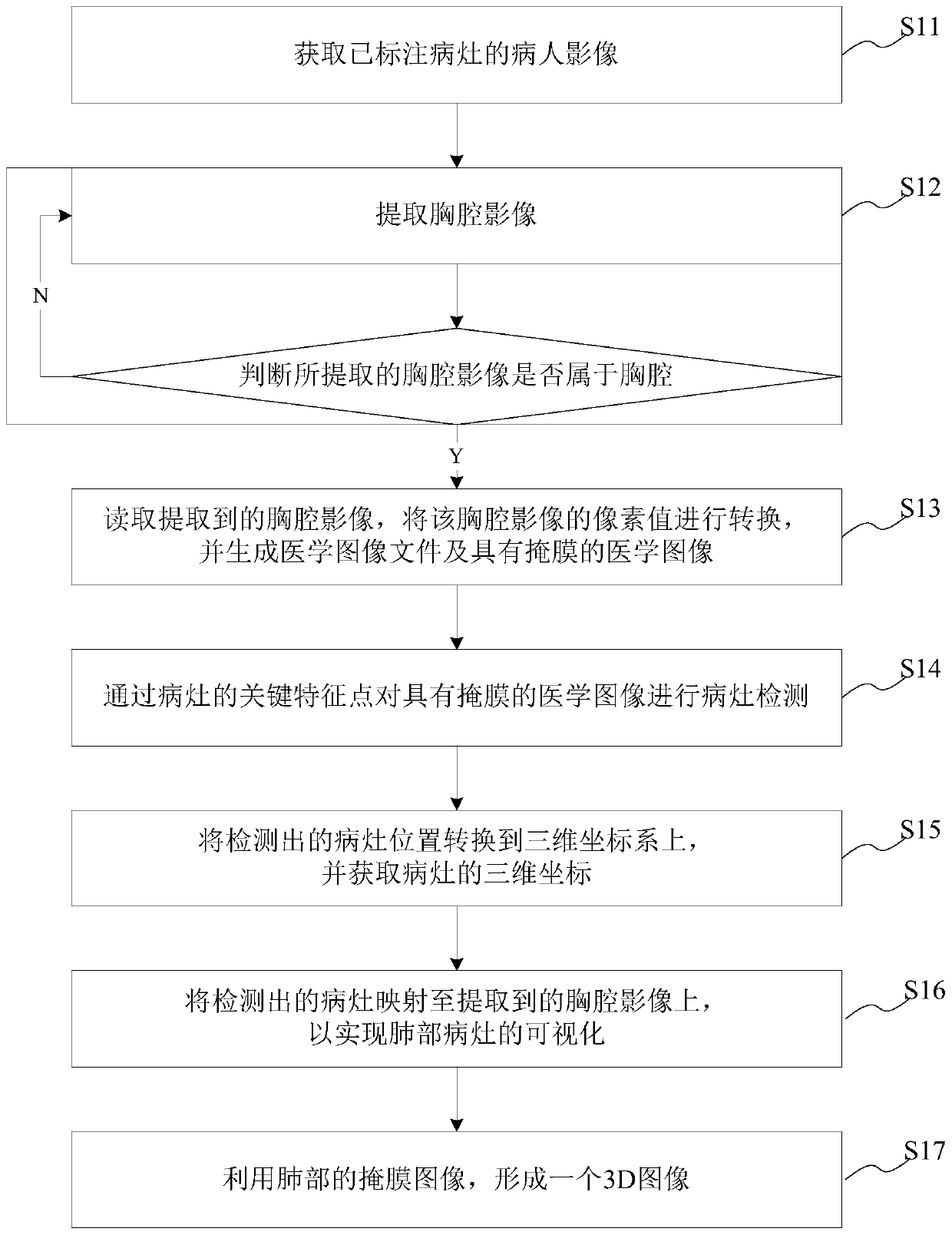
[0032] This embodiment provides a method for detecting lung lesions in images, including:

[0033] Obtain a patient image with marked lesions; identify the marked area outline from the marked lesion image of the patient, and train the identified area outline to obtain key feature points for predicting lesions;

[0034] Extract the chest image from the patient's whole body image, and determine whether the extracted chest image belongs to the chest; if yes, perform the next step; if not, re-extract the chest image;

[0035] Read the extracted thoracic image, convert the pixel value of the thoracic image, and generate a medical image file and a medical image with a mask;

[0036] Lesion detection is performed on the medical image with a mask through the key feature points of the lesion;

[0037] Map the detected lesions to the extracted thoracic images to visualize the lung lesions, form a 3D model of the lungs, and output them.

[0038] The method for detecting lung lesions in...

Embodiment 2

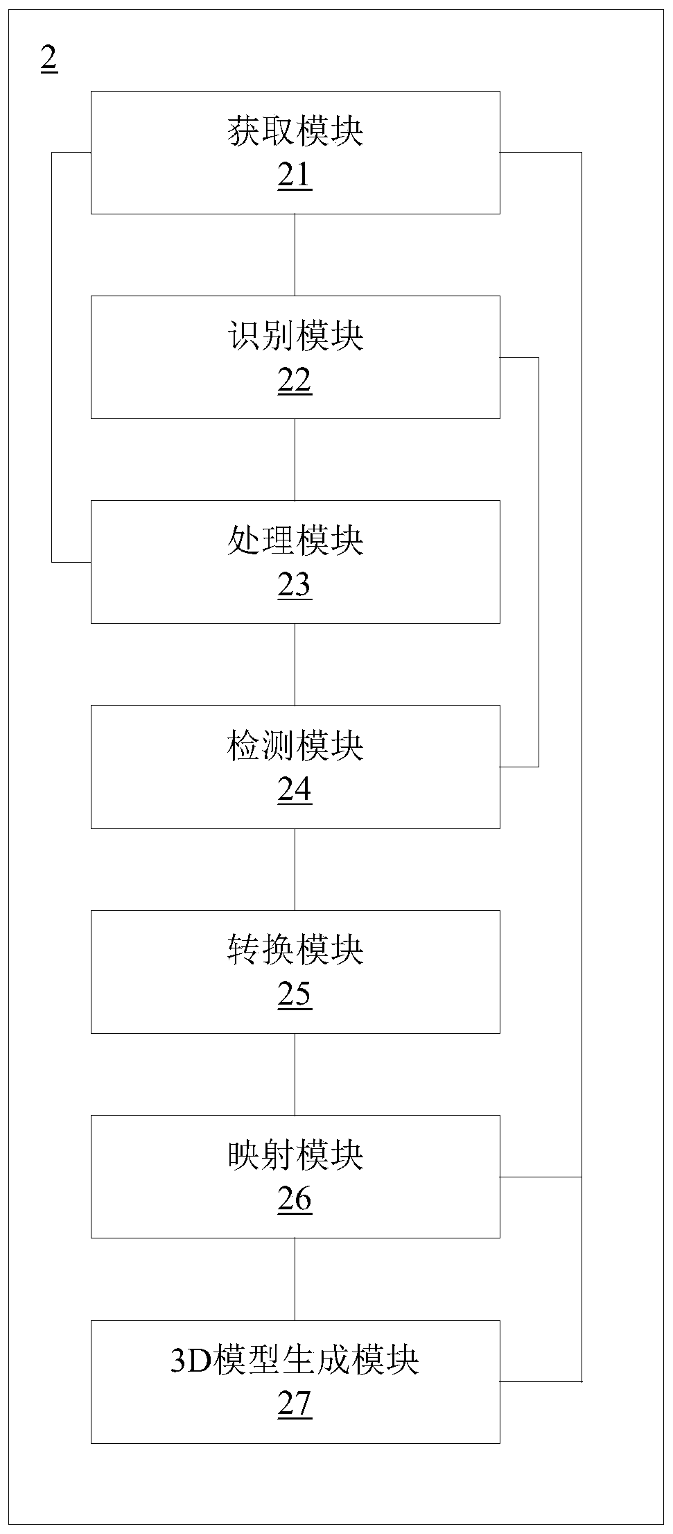
[0077] The present embodiment provides a detection system for pulmonary lesions, including:

[0078] An acquisition module, configured to acquire images of patients with marked lesions;

[0079] The recognition module is used to recognize the contour of the marked area from the image of the patient with the marked lesion, and train the contour of the recognized area to obtain key feature points for predicting the lesion;

[0080] The processing module is used to extract the chest cavity image from the patient's whole body image, and judge whether the extracted chest cavity image belongs to the chest cavity; if so, read the extracted chest cavity image, convert the pixel values ​​of the chest cavity image, and generate a medical image file and medical image with mask; if not, re-extract chest image;

[0081] The detection module is used to detect the focus of the medical image with the mask through the key feature points of the focus;

[0082] The mapping module is used to ma...

Embodiment 3

[0112] This embodiment provides a terminal, including: a processor, a memory, a transceiver, a communication interface or / and a system bus; the memory and the communication interface are connected to the processor and the transceiver through the system bus to complete mutual communication, and the memory is used for The computer program is stored, the communication interface is used to communicate with other devices, the processor and the transceiver are used to run the computer program, so that the device executes the steps of the method for detecting lung lesions above.

[0113] The system bus mentioned above may be a Peripheral Component Interconnect (PCI for short) bus or an Extended Industry Standard Architecture (EISA for short) bus or the like. The system bus can be divided into address bus, data bus, control bus and so on. The communication interface is used to realize the communication between the database access device and other devices (such as client, read-write li...


PUM


Abstract

The invention provides a lung lesion detection method and system, a storage medium, a terminal and a display system. The lung lesion detection method comprises the steps of acquiring a patient image marked with a lesion; identifying the marked area contour, and training the identified area contour to obtain key feature points; extracting a thoracic cavity image from the whole body image of the patient, and judging whether the extracted thoracic cavity image belongs to the thoracic cavity or not; if so, executing the next step; if not, re-extracting the thoracic cavity image; reading the extracted thoracic cavity image, converting pixel values of the thoracic cavity image, and genertaing a medical image file and a medical image with a mask; performing focus detection on the medical image with the mask through the key feature points of the focus; and mapping the detected lesions to the extracted thoracic cavity image to realize visualization of the lung lesion and form a lung 3D model. Amedical auxiliary means is provided for doctors, and the doctors are assisted to accurately diagnose the position of the lung focus in the image.

Description

technical field [0001] The invention belongs to the technical field of medical image processing, and relates to a detection method and system, in particular to a detection method, system, storage medium, terminal and display system for lung lesions. Background technique [0002] The lungs are an important respiratory organ of the human body and also undertake the function of hematopoiesis. The lungs are connected to the trachea, bronchi, throat, and nose of the human body, and any problem in any of them will affect our respiratory system. So, what does it mean to have lesions in the lungs? In fact, this suggests that there is a disease in our lungs, which may be pneumonia, pulmonary nodules, pulmonary fibrosis and other problems. However, in the prior art, a doctor judges lesions on a medical image with naked eyes, resulting in inaccurate detection. [0003] Therefore, how to provide a detection method, system, storage medium, terminal and display system for lung lesions,...

Claims


Application Information

Patent Timeline
IPC IPC(8): G06T7/00G06T7/62G06T17/00G16H30/20G16H30/40
CPCG06T7/0012G06T7/62G06T17/00G06T2207/10081G06T2207/20081G06T2207/20084G06T2207/30064G16H30/20G16H30/40
Inventor 李超李萍华涛管一晖
Owner SHANGHAI ENG RES CENT FOR BROADBAND TECH & APPL
Why Eureka
Try Eureka