
3D printing device and method of multi-scale fiber-reinforced micro-channel active tubular tissue

A fiber-reinforced, printing device technology, applied in tissue cell/virus culture devices, biomaterial sampling methods, biochemical equipment and methods, etc., can solve the problem that hydrogel tubular tissues cannot meet the mechanical properties of tubular tissues. , The problem of nutrient supply and delivery of three-dimensional tubular tissue has not been well solved, so as to achieve the effect of promoting survival and growth and ensuring supply.

Active Publication Date: 2019-07-12
XI AN JIAOTONG UNIV
View PDF4 Cites 11 Cited by
  • Summary
  • Abstract
  • Description
  • Claims
  • Application Information

AI Technical Summary

Problems solved by technology

However, there are still some problems in the tubular tissue constructed by the existing methods: (1) the hydrogel tubular tissue constructed by cell printing cannot meet the mechanical properties requirements of tubular tissue in different parts of the human body; (2) the pure hydrogel tubular structure It can't simulate the real environment with dense micro-nano fibers in the body; (3) The problem of nutrient supply and transportation in three-dimensional tubular tissue has not been well solved

Method used


Image

  • 3D printing device and method of multi-scale fiber-reinforced micro-channel active tubular tissue
  • 3D printing device and method of multi-scale fiber-reinforced micro-channel active tubular tissue
  • 3D printing device and method of multi-scale fiber-reinforced micro-channel active tubular tissue

Examples


Embodiment Construction

[0026] The present invention will be described in further detail below in conjunction with the accompanying drawings and embodiments.

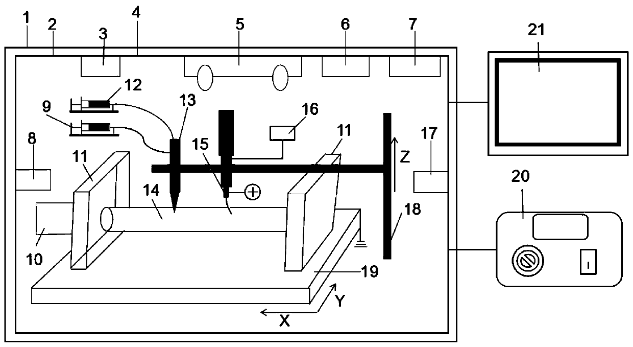
[0027] refer to figure 1 , a multi-scale fiber-reinforced microfluidic channel active tubular tissue 3D printing device, including an ultra-clean test bench 1, a printing device shell 2 is placed inside the ultra-clean test bench 1, and a printing platform 19 is arranged in the printing device shell 2, and a printing platform 19 Grounded, the printing platform 19 can move along the X-Y two directions, the receiving cylinder 14 is connected with the bearing seat 11 on the printing platform 19, the receiving cylinder 14 is installed on the bearing seat 11, and it is easy to disassemble; the receiving cylinder 14 moves with the printing platform 19 , can realize horizontal movement in XY two directions; the end of the receiving drum 14 is connected to the output shaft of the motor 10, and the receiving drum 14 can rotate at a certain speed under ...


PUM


Abstract

The invention relates to a 3D printing device and method of a multi-scale fiber-reinforced micro-channel active tubular tissue, wherein the device comprises a printing device shell, a printing platform is arranged in the printing device shell, the printing platform can move along the X-Y direction, a receiving roller is connected with the printing platform, the receiving roller is connected with amotor, a coaxial spray nozzle and a melting electrostatic printing device are arranged above the receiving roller, the coaxial spray nozzle and the melting electrostatic printing device are both fixed on a Z-axis moving platform, an inner spray nozzle and an outer spray nozzle of the coaxial spray nozzle are connected with a precise injector respectively. Through the combination of the melting electrostatic printing technology, melt extrusion molding technology and cell printing technology, the on-demand adjustment of three-dimensional tubular tissue mechanical properties can be realized, microchannels are formed in the annular wall of the tubular tissue, nutrients and metabolic wastes are transferred to cells in the tissue in time, the living environment with densely covered fibers in the body can be simulated more realistically, and the expression of cell function is facilitated.

Description

technical field [0001] The invention relates to the field of biological manufacturing and biological 3D printing, in particular to a device and method for 3D printing of multi-scale fiber-reinforced microfluidic active tubular tissue. Background technique [0002] Cell printing is based on the principle and basis of traditional additive manufacturing technology, using biological materials, active cells, growth factors, etc. Organ fabrication provides a new technical means. Among them, the use of cell printing technology to construct three-dimensional tubular structures to simulate tubular tissues such as human urinary tract, intestine, esophagus, trachea, bile duct, and blood vessels has attracted widespread attention from scholars at home and abroad. However, there are still some problems in the tubular tissue constructed by the existing method: (1) the hydrogel tubular tissue constructed by cell printing cannot meet the mechanical properties requirements of tubular tissue...

Claims


Application Information

Patent Timeline
IPC IPC(8): C12M3/00B33Y30/00
CPCB33Y30/00C12M33/00
Inventor 贺健康梁红涛李涤尘
Owner XI AN JIAOTONG UNIV
Learn More