Method and device for determining tubular tissue region of interest, equipment and medium

A region of interest, tubular technology, applied in the field of medical imaging, can solve the problems of time-consuming and labor-intensive editing, and the difficulty of determining the outer wall of blood vessels, etc., to achieve the effects of time-consuming and labor-intensive, accurate measurement and size assessment, and simple determination methods

Active Publication Date: 2019-10-18
SHANGHAI UNITED IMAGING HEALTHCARE
View PDF3 Cites 6 Cited by
  • Summary
  • Abstract
  • Description
  • Claims
  • Application Information

AI Technical Summary

Problems solved by technology

[0004] The problem with this type of vascular plaque detection method at present is that the outer wall of the blood vessel is very difficult to determine, and doctors often need to edit the inner and outer contours of the blood vessel on the cross section, especially when the blood vessel is long and the cross section taken along the centerline The number is very large, and editing is time-consuming and laborious

Method used


Image

  • Method and device for determining tubular tissue region of interest, equipment and medium
  • Method and device for determining tubular tissue region of interest, equipment and medium
  • Method and device for determining tubular tissue region of interest, equipment and medium

Examples


Embodiment 1

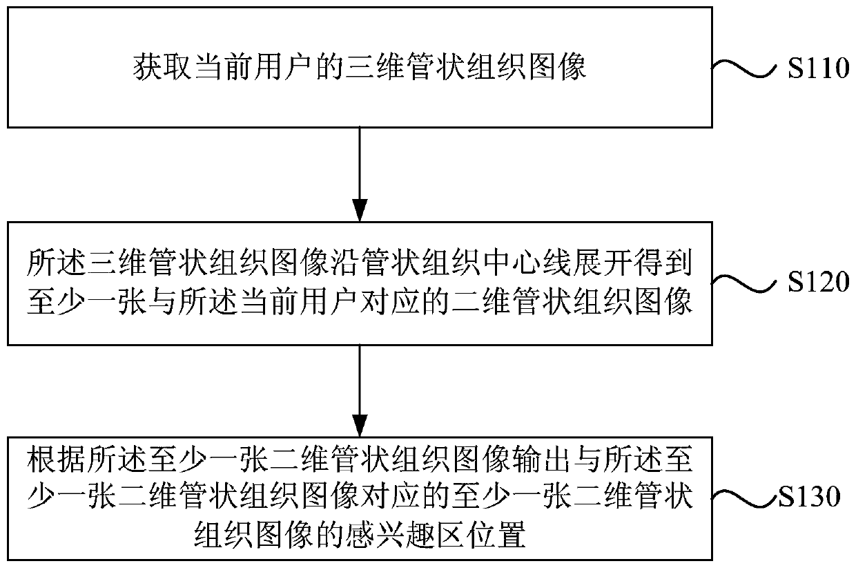
[0052] figure 1 It is a flowchart of a method for determining a region of interest in tubular tissue provided by Embodiment 1 of the present invention. This embodiment is applicable to the situation of determining a region of interest in tubular tissue. The method can be determined by determining the region of interest in tubular tissue The device is implemented, which specifically includes the following steps:

[0053] S110. Acquire a three-dimensional tubular tissue image of the current user.

[0054]Among them, tubular tissues include blood vessels and trachea. The three-dimensional tubular tissue image may include enhanced CT blood vessel images, may also include images obtained by MR blood vessel imaging such as MR angiography images or MR TOF images, and any other three-dimensional images that can image blood vessels.

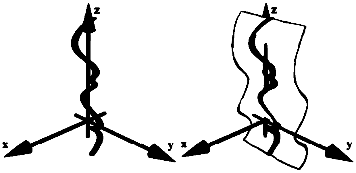
[0055] S120. Expand the three-dimensional tubular tissue image along the centerline of the tubular tissue to obtain at least one two-dimensional tubula...

Embodiment 2

[0064] Figure 4 It is a flowchart of a method for determining a region of interest in tubular tissue provided by Embodiment 2 of the present invention. This embodiment is optimized based on the above embodiments. In this embodiment, the three-dimensional tubular tissue image described in the step is expanded along the tubular tissue centerline to obtain at least one two-dimensional tubular tissue image corresponding to the current user for further optimization. It is: unfolding the three-dimensional tubular tissue image along the centerline of the current user's tubular tissue to obtain at least one two-dimensional tubular tissue image corresponding to the current user from different angles. On this basis, the step of outputting the ROI position of the at least one two-dimensional tubular tissue image corresponding to the at least one two-dimensional tubular tissue image according to the at least one two-dimensional tubular tissue image is further optimized as follows: the at...

Embodiment 3

[0078] Figure 5 It is a schematic diagram of an optional embodiment of a method for determining a region of interest in tubular tissue provided by Embodiment 3 of the present invention. On the basis of the foregoing embodiments, a preferred embodiment is provided. The method for determining a region of interest in tubular tissue includes:

[0079] Acquire historical 3D tubular tissue images.

[0080] The centerline of the historical three-dimensional tubular tissue is extracted by means of automatic extraction, semi-automatic extraction or manual extraction, etc. to extract the centerline of the tubular tissue.

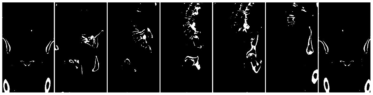
[0081] Expand the historical 3D tubular tissue image along the tubular tissue centerline to obtain at least one 2D tubular tissue image corresponding to the historical 3D tubular tissue image at different angles, which is convenient for intuitive observation. After unfolding, the same tubular tissue image according to different reconstruction angles can be obtained...


PUM


Abstract

The embodiment of the invention discloses a method and a device for determining a tubular tissue region of interest, equipment and a medium. The method comprises the following steps: acquiring a three-dimensional tubular tissue image of a current user; wherein the three-dimensional tubular tissue image is expanded along a tubular tissue center line to obtain at least one two-dimensional tubular tissue image corresponding to the current user; and outputting a region of interest position of at least one two-dimensional tubular tissue image corresponding to the at least one two-dimensional tubular tissue image according to the at least one two-dimensional tubular tissue image. According to the embodiment of the invention, through obtaining a three-dimensional tubular tissue image of a currentuser, and after at least one two-dimensional tubular tissue image is obtained by expanding along the central line of the tubular tissue, the two-dimensional tubular tissue image is input into a pre-trained classifier to output the position of the region of interest of the corresponding two-dimensional tubular tissue image, so that the determination method of the region of interest of the tubulartissue is simpler, and the measurement and size evaluation of the region of interest of the tubular tissue are more accurate.

Description

technical field [0001] Embodiments of the present invention relate to the technical field of medical imaging, and in particular to a method, device, device and medium for determining a region of interest in tubular tissue. Background technique [0002] Tubular tissue is the vertical and horizontal connection of cells with the same nature in higher plants, and the partition wall between cells disappears, and the thin tubular tissue formed by interpenetration is the passage of water, such as blood vessels and trachea. The detection of vascular diseases in medical images has always been a topic of great concern to medicine, especially for coronary artery calcification and plaque detection, which is of great significance to the diagnosis of heart disease. [0003] Taking blood vessels as an example, in the past few years, with the development and maturity of vascular imaging technology, especially after the heart 64-slice CT gradually occupied the market, many plaque detection t...

Claims


Application Information

Patent Timeline
Patent Type & Authority Applications(China)
IPC IPC(8): G06T7/00G16H30/20
CPCG06T7/0012G16H30/20G06T2207/20104G06T2207/20081G06T2207/30101
Inventor 王晓东
Owner SHANGHAI UNITED IMAGING HEALTHCARE
Why Eureka
Try Eureka