
A Method for Enhancing the Contrast of Early Embryo Optical Coherence Tomography

A technology of optical coherence tomography and image contrast, applied in the field of biomedical imaging, can solve the problems of expensive imaging system and limited applicability of mammalian embryonic vascular reconstruction

Active Publication Date: 2020-11-13
XIAMEN UNIV
View PDF0 Cites 0 Cited by
  • Summary
  • Abstract
  • Description
  • Claims
  • Application Information

AI Technical Summary

Problems solved by technology

Larin et al. used swept-frequency optical coherence tomography to study the normal vascular development of zebrafish and chicken embryos, but the applicability to mammalian embryo vascular remodeling is limited, and the imaging system is expensive

Method used


Image

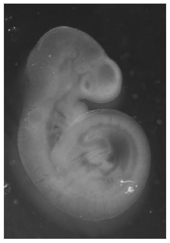
  • A Method for Enhancing the Contrast of Early Embryo Optical Coherence Tomography
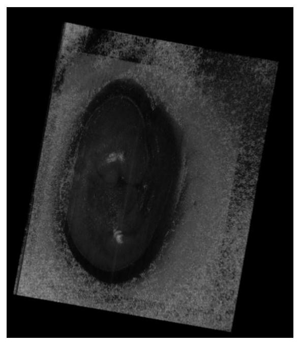
  • A Method for Enhancing the Contrast of Early Embryo Optical Coherence Tomography
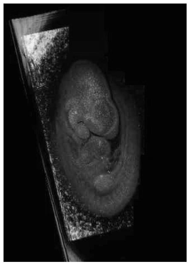
  • A Method for Enhancing the Contrast of Early Embryo Optical Coherence Tomography

Examples


Embodiment 1

[0029] A method for enhancing the contrast of early embryo optical coherence tomography image includes the following steps:

[0030] (1) Separate early embryos with intact placenta and vitelline membrane from the mother, as follows:

[0031] A. According to the pregnancy date marked on the cage, the mice with gestational age of 9.5 days (E9.5) and 10.5 days (E10.5) were selected accurately and their necks were sacrificed. Carefully disinfect the surface of the abdomen with 75% ethanol. Use high-pressure steam sterilized ophthalmological forceps to lift the abdominal skin, use ophthalmic scissors to make an incision on the midline of the abdomen and open the skin to both sides, continue to open the entire abdominal cavity, gently move the organs to see the beaded embryos "V"-shaped uterus, cut off the cervix, and place several embryos in a 55mm petri dish containing DMEM medium. Use sterile ophthalmic scissors to cut the uterus along the opposite side of the mesangium or slowly te...


PUM


Abstract

The invention discloses a method for enhancing the contrast of an early embryo optical coherence tomography image, which comprises the following steps: (1) separating an early embryo with a complete placenta and vitelline membrane from a mother; (2) placing the early embryo in DMEM In the embryo in vitro culture medium of culture medium, serum, penicillin, streptomycin, non-essential amino acid and glutamic acid, cultivate 3-8h, contain nano gold rod in this embryo in vitro culture medium; (3) step (2) After the cultured early embryos were stripped of the vitelline membrane, placenta and amniotic membrane, they were sequentially photographed with an optical microscope, OCT imaging to obtain two-dimensional images, and 3D scanning to obtain three-dimensional images. The present invention uses gold nanorods as an optical contrast agent to improve the performance of traditional SD-OCT imaging of early embryos, improve the quality of imaging, and realize 3D visual imaging, which has very important guiding significance for guiding clinical embryonic development and improves Important experimental data references and research strategies.

Description

Technical field [0001] The invention belongs to the technical field of biomedical imaging, and specifically relates to a method for enhancing the contrast of early embryo optical coherence tomography images. Background technique [0002] At present, there are still many problems in the study of mammalian embryonic development after implantation, such as structural morphology problems. During development, cells are not arranged in a disorderly manner. Although they have multidirectional differentiation potential, they do not migrate and distribute randomly. Instead, they differentiate into specific cell types with different functions, forming complex tissues and organs with different shapes and functions. Tissues and organs are arranged in a certain way to form a complete system. This complete organ formation process is very complicated. To achieve early and continuous observation of embryonic development and imaging in a living state is currently an important clinical research f...

Claims


Application Information

Patent Timeline
Patent Type & Authority Patents(China)
IPC IPC(8): C12Q1/02G01N21/64C12N5/073B22F9/24B22F1/00
CPCG01N21/6458G01N21/6486C12N5/0604B22F9/24B22F1/07B22F1/054
Inventor 赵庆亮黄雅丽刘刚黄豆豆张小芬林文珍刘基嫣严文豪龚向莲
Owner XIAMEN UNIV
Learn More