Hip joint X-ray image segmentation method and system based on local vision clue

A technology of visual cues and light images, applied in the field of deep learning and computer vision, can solve limited problems, achieve the effect of reducing complexity, model robustness, and improving model effect

Inactive Publication Date: 2019-11-22
CHONGQING UNIV OF POSTS & TELECOMM
View PDF0 Cites 7 Cited by
  • Summary
  • Abstract
  • Description
  • Claims
  • Application Information

AI Technical Summary

Problems solved by technology

However, the classic full convolution segmentation algorithm requires a large amount of accur

Method used


Image

  • Hip joint X-ray image segmentation method and system based on local vision clue
  • Hip joint X-ray image segmentation method and system based on local vision clue
  • Hip joint X-ray image segmentation method and system based on local vision clue

Examples


Example Embodiment

[0028] The present invention will be further described below in conjunction with the accompanying drawings of the specification.

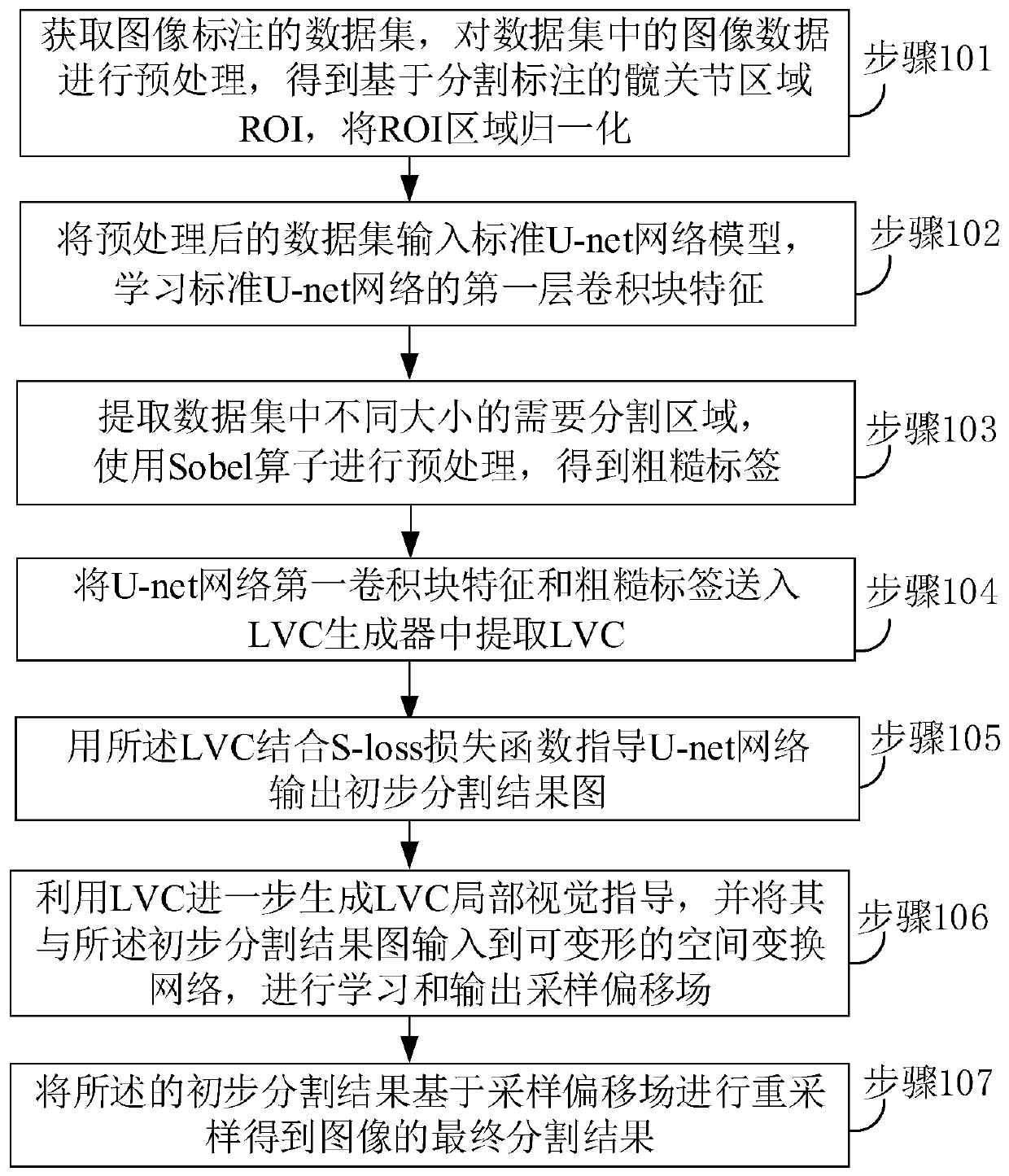
[0029] figure 1 It is a schematic flowchart of a hip joint X-ray image segmentation method based on local visual cues provided by an embodiment of the present invention. The method includes the following steps:

[0030] Step 101: Obtain a data set labeled by X-ray images, preprocess the image data in the data set to obtain a hip joint region (ROI) based on segmentation and labeling, and normalize the ROI region;

[0031] Specifically, when preprocessing the image data in the data set, you can first use the labelme annotation tool to obtain the X-ray image annotation data set, convert the data set format to COCO2014 format, and use the pycocotools toolkit to deserialize the json to obtain the network Input, recalculate the ROI area according to the range of the divided points and random numbers, enhance the data, and normalize it to 128x128.

[0032] Step 1...


PUM


Abstract

The invention relates to a hip joint X-ray image segmentation method and system based on a local vision clue. The method comprises the following steps: preprocessing the image data in the data set; learning a first-layer convolution block feature of the standard U-net network; obtaining a rough label by using a Sobel operator; extracting a local visual clue LVC through the first convolution blockfeature and the rough label; combining LVC with an S-loss loss function to guide a U-net network to output a preliminary segmentation result graph; utilizing LVC to generate LVC local visual guidance,outputting a sampling offset field with the preliminary segmentation result image through a deformable spatial transformation network, and resampling the preliminary segmentation result image to obtain a final segmentation result of the image. According to the method, the problem that a supervised learning model is easily influenced by label noise in the medical image segmentation process is solved, and the complexity of medical image label generation is reduced.

Description

technical field [0001] The invention belongs to the technical field of deep learning and computer vision. Specifically, it relates to a weakly supervised hip image segmentation method based on local visual cues to automatically correct semantic segmentation errors. Background technique [0002] Convolutional neural networks have achieved very good results in the field of computer vision and are excellent image feature extractors for various image tasks. However, computerized tomography CT images, magnetic resonance imaging MRI images, and X-ray (X-ray) images in the medical field are very different from natural images due to the difficulty in data labeling and small amount of data, which leads to convolutional neural networks. It cannot be well applied to medical images. [0003] In the medical field of hip dysplasia, the screening of developmental dysplasia of the hip (DDH) mainly relies on the doctor's geometric angle calculation and observation. For hip joint segmentat...

Claims


Application Information

Patent Timeline
IPC IPC(8): G06T7/11
CPCG06T7/11G06T2207/10116G06T2207/20081G06T2207/20084G06T2207/30008
Inventor 舒禹程李伟生吴潇马旭齐大逊赵君
Owner CHONGQING UNIV OF POSTS & TELECOMM
Why Eureka
Try Eureka