Diagnostic imaging hepatic tissue-specific contrast medium comprising manganese silicate nanoparticle

A nanoparticle, liver tissue technology, applied in MRI/MRI contrast agent, nanomedicine, nanotechnology, etc., can solve the problems of unresearched differential diagnosis, slow speed of Mn2+, etc.

Active Publication Date: 2019-11-26
POSTECH ACAD IND FOUND +1
View PDF1 Cites 2 Cited by
  • Summary
  • Abstract
  • Description
  • Claims
  • Application Information

AI Technical Summary

Problems solved by technology

However, due to Mn released from Kupffer cells 2+ The speed is slow, so it has the disadvantage of relatively long time to obtain images of liver cancer, and the possibility of using it for differential diagnosis according to the characteristics of liver cancer has not been studied

Method used


Image

  • Diagnostic imaging hepatic tissue-specific contrast medium comprising manganese silicate nanoparticle
  • Diagnostic imaging hepatic tissue-specific contrast medium comprising manganese silicate nanoparticle
  • Diagnostic imaging hepatic tissue-specific contrast medium comprising manganese silicate nanoparticle

Examples


example 1

[0109] Example 1. Material preparation and steps

[0110] 1-1. Experimental materials

[0111] Use purchased MnCl without any purification 2 4H 2 O(kanto), sodium oleate (TCI), 1-octadecene (Aldrich), CO-520 (Aldrich), tetraethyl silicate (Acros), NH 4 OH(Samchun Chem.), Ni(NO 3 )6H 2 O(Strem), Cu(NO 3 ) 2 ·3H 2 O(Strem), NaBH 4 (Samchun Chem.), 2-[methoxy(polyethyleneoxy)-propyl]9-12-trimethoxysilane (MPEOPS, Gelest, Inc.), fluorescein isothiocyanate (FITC, Aldrich) and 3-aminopropyltriethoxysilane (APTES, Aldrich).

[0112] 1-2. Preparation of HMS (hollow manganese silicate)

[0113] According to previously reported techniques, by making MnO@SiO 2 / Cu 2+ Annealing followed by selective etching of the outer silica shell of Cu@HSNPs (hollow-structured NPs) (Kim, J.G., Kim, S.M. & Lee, I.S. Mechanistic insight into the yolk@shell transformation of MnO@silica nanospheres incorporating Ni 2+ ionstoward a colloidal hollow nanoreactor.Small 11, 1930-1938(2015)) ...

example 2

[0128] Example 2. Mn Relaxation Properties and T1 Contrast Enhancement Properties of Hollow Manganese Silicate (HMS) Nanoparticles

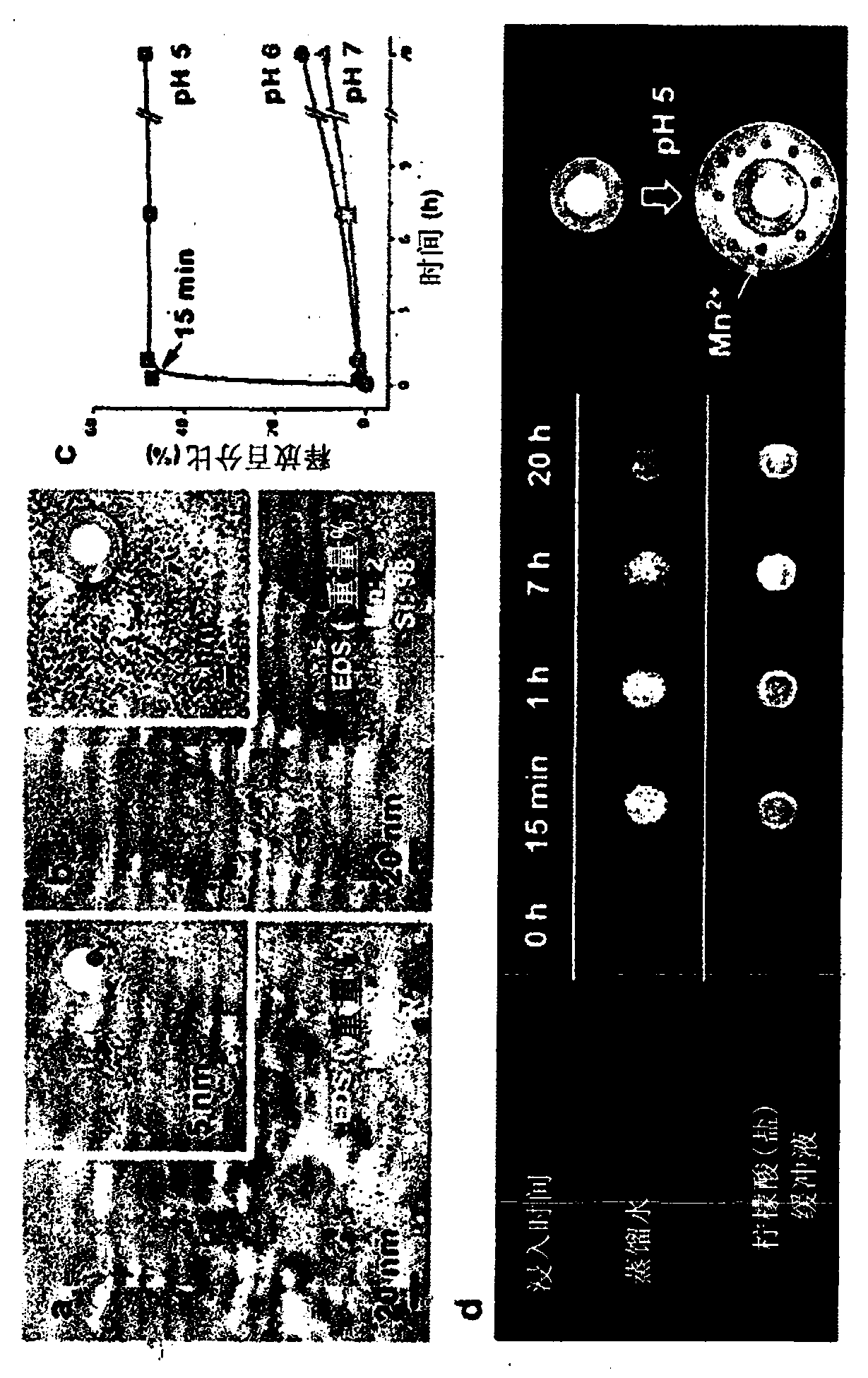
[0129] The Mn relaxation properties and T1 contrast enhancement properties of the HMS prepared in Examples 1-2 were tested according to Examples 1-3. TEM was used to observe the synthesized nanoparticles and the nanoparticles isolated from the pH 5.0 buffer solution ( figure 1 a and figure 1 b).

[0130] like figure 1 c and figure 1 The result of d, when the PEG-modified HMS containing 19.3wt% Mn was immersed in the buffer solution of pH 5.0, all the Mn that could be released 2+ Immediate release and increase to free Mn within 15 minutes 2+ the maximum concentration of ions. On the contrary, when the PEG-modified HMS was immersed in a pH 6.0 buffer solution or distilled water, Mn 2+ Ions are released very slowly ( figure 1 c). Additionally, T1-weighted images in distilled water were not affected by HMS. However, when HMS was added to th...

example 3

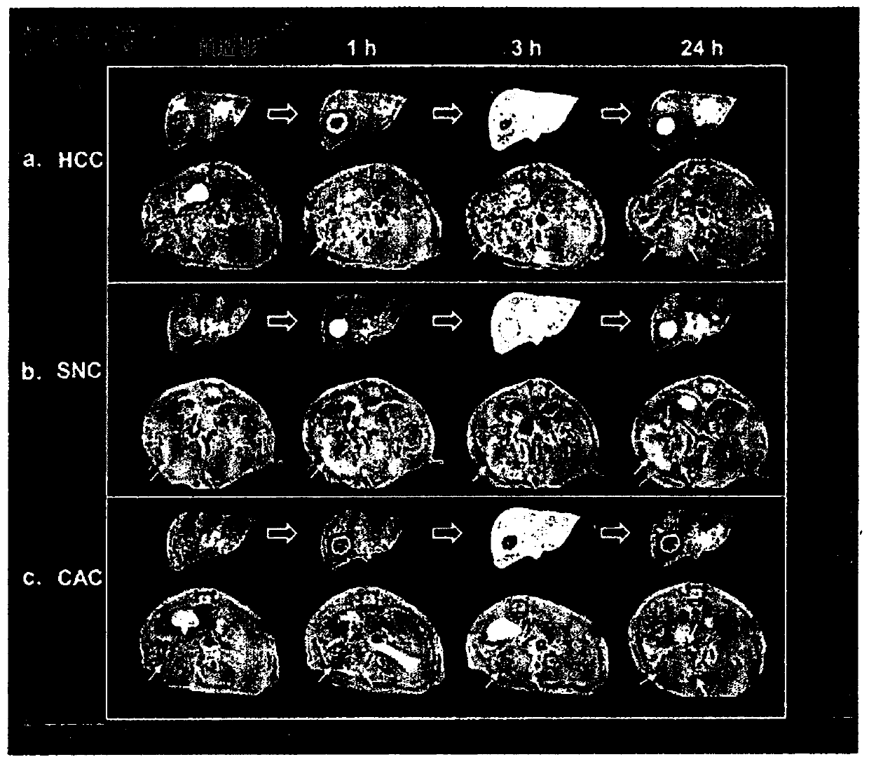
[0132] Example 3. Evaluation of contrast-enhancing patterns of HMS in HCC

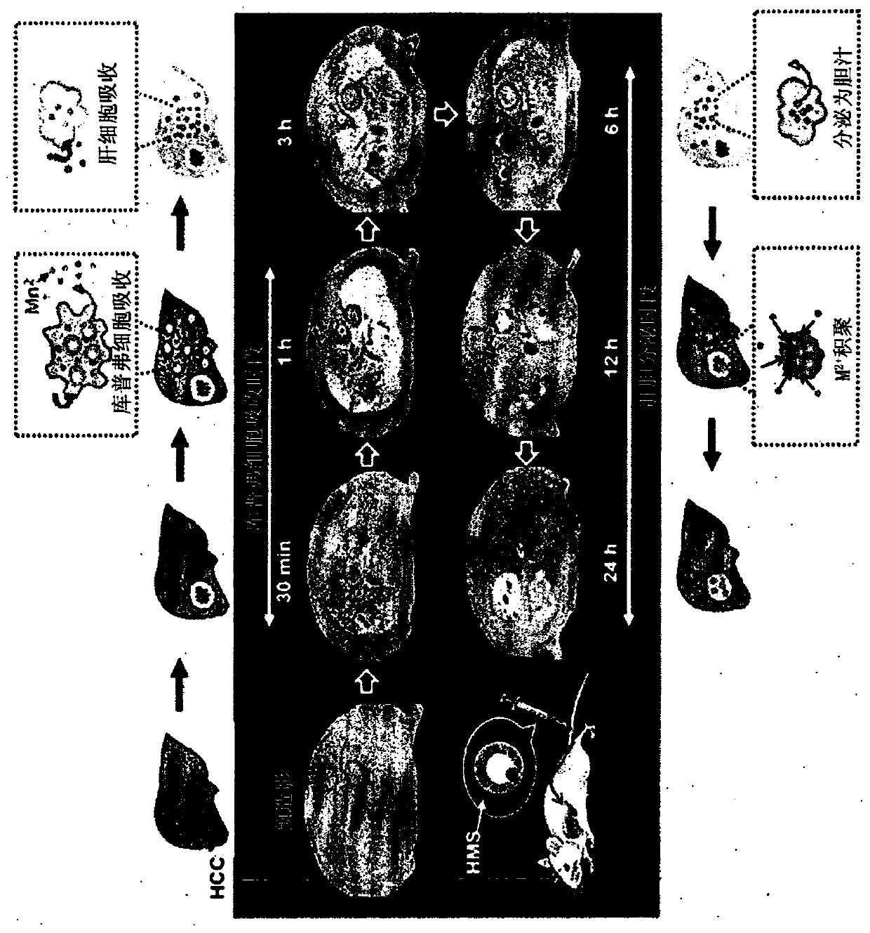
[0133] First, HMS was injected (3 mg / kg body weight) into a human hepatocellular carcinoma (HCC) model and observed within 0.5 hours to 24 hours after HMS injection to evaluate the potential of HMS as a contrast agent for distinguishing liver tumors sex.

[0134] like figure 2 As a result, obvious contrast enhancement was observed within 0.5 hours to 6 hours after HMS injection, and then slowly began to brighten from the peripheral area to the central area within 6 hours to 24 hours, and the necrotic area was not enhanced.

[0135] It can be interpreted that these results show that during the initial 0.5 h to 6 h period (Cooper cell (Coopercell) uptake phase), Mn 2+ Ions are released from nanoparticles absorbed in Kupffer cells and then released to Mn in liver tissue within 6 hours to 24 hours (biliary release period) 2+ Ions are taken up by hepatocytes and released into bile, ie, ions are taken up...


PUM


Abstract

The present invention relates to a diagnostic imaging manganese silicate hepatic tumor-specific MRI contrast medium which releases manganese ions (Mn2+) in an acidic condition and a method for characterizing a hepatic tissue, using the same. Exhibiting different patterns of T1-weighted imaging including vascularity, cell density, mitochondrial activity, hepatocellular affinity, etc., according totissue specificity of normal hepatic tissues and diseased liver tissues (particularly hepatic tumor), within relatively short period of time, the present invention allows disease-specific characteristics of hepatic tumor to be detected at an appropriate time through the analysis of such T1-weighted imaging. Particularly, the present invention is expected to be very effectively used in discerning and diagnosing kinds of hepatic tumor or in determining therapeutic effects and further to be helpful in diagnosing diseases generated in organs other than the liver according to tissue specificity.

Description

technical field [0001] The present invention relates to a contrast agent for liver tissue imaging diagnosis, more particularly, to a contrast agent comprising releasing manganese ions (Mn 2+ ) manganese silicate contrast agent for specific imaging diagnosis of liver cancer, or more specifically, relates to an MRI (magnetic resonance imaging) contrast agent for specific imaging diagnosis of liver tumors. Background technique [0002] The role of contrast agents in magnetic resonance imaging (MRI) is to improve the ability of diagnosis or differential diagnosis by enhancing the ability to detect lesions or to characterize lesions by contrast enhancement of organs or lesions. Currently, because the paramagnetic Gd 3+ Chelate-based contrast agents are non-specific extracellular fluid (ECF) contrast agents based on differences in vascular distribution, so paramagnetic Gd 3+ Chelate contrast agents are widely used in liver MRI. Among them, Gd-EOB-DTPA, which is called a liver-s...

Claims


Application Information

Patent Timeline
Patent Type & Authority Applications(China)
IPC IPC(8): A61K49/18
CPCA61K49/1818A61K49/08A61K49/1824A61K49/1827A61K49/183B82Y5/00
Inventor 李源宰李贞姬张文瑄李寅洙金晋究
Owner POSTECH ACAD IND FOUND
Why Eureka
Try Eureka