

Lung tumor automatic sketching method based on deep learning
A deep learning and tumor technology, which is applied in the field of medical image processing technology and deep learning, can solve the problems of low tumor delineation accuracy, inaccurate manual delineation, time-consuming and labor-intensive problems, so as to improve the safety process of surgery, reduce the workload and improve the delineation efficiency Effect
Patent Information
- Authority / Receiving Office
- CN · China
- Current Assignee / Owner
- Publication Date
- 2020-01-17
Smart Images
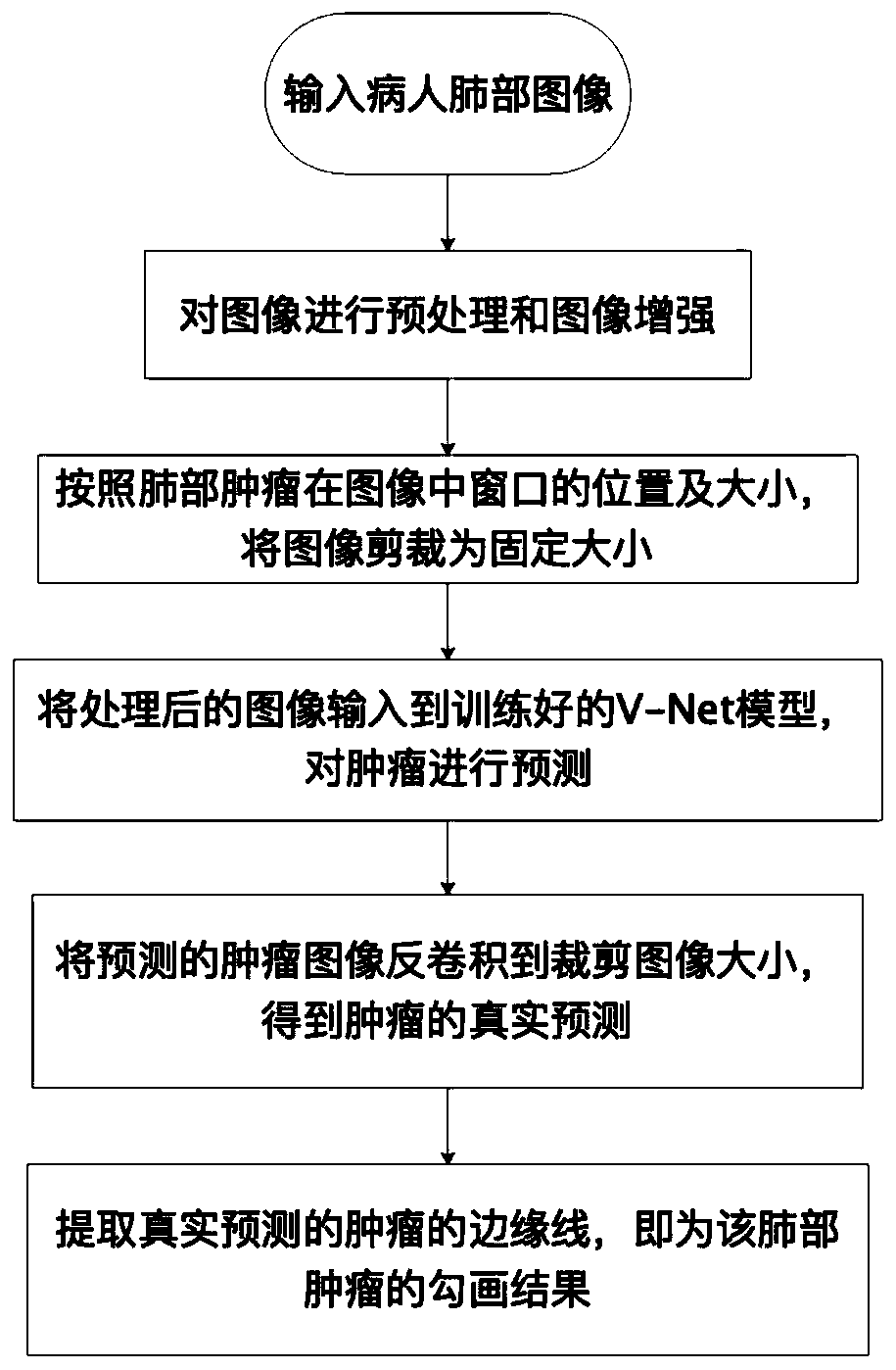
Figure 1 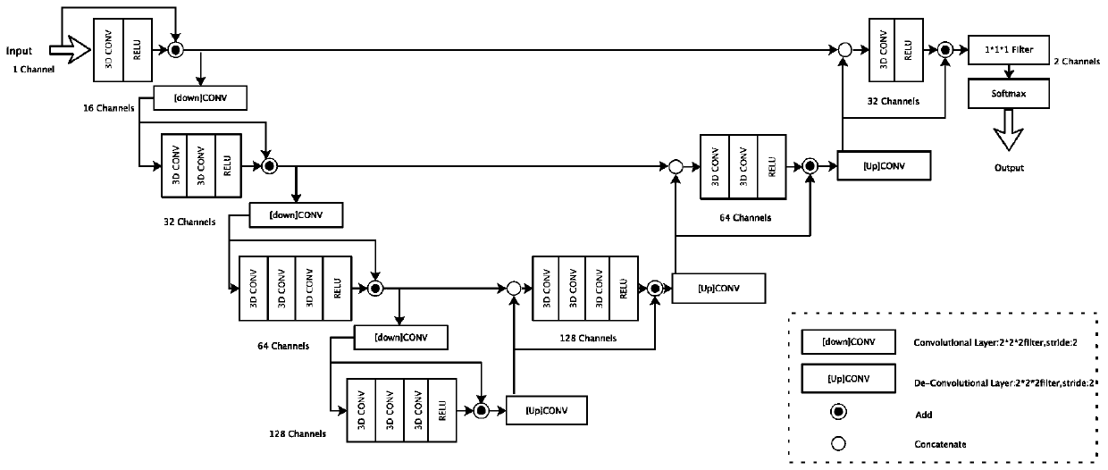
Figure 2 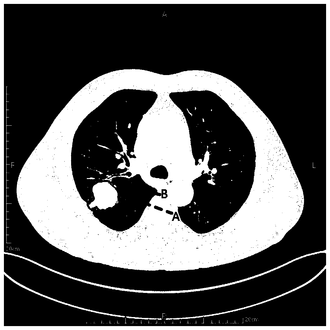
Figure 3
Abstract
Description
technical field
[0001] The invention relates to the fields of medical image processing technology and deep learning, in particular to an automatic delineation method for lung tumors based on deep learning. Background technique
[0002] Lung cancer is one of the malignant tumors that seriously threaten human health, and its death rate ranks first in cancer. There are about 1.8 million new lung cancer cases (accounting for 13% of all tumors) and 1.6 million deaths (accounting for 19.4% of all tumors) in the world every year. The annual survival rate is only 18%. If diagnosed early, the 5-year survival rate of lung cancer patients can increase to 70%, improving the prognosis of patients.
[0003] Modern medical technology is developing day by day, a large part of which is due to the maturity of medical imaging technology. Including CT technology, MRI technology, etc. These technologies help doctors understand the internal pathological structure of patients and formulate preci...
Examples
Embodiment Construction
[0023] In order to make the purpose, technical solution and advantages of the present invention clearer, the implementation manners of the present invention will be further described in detail below.
[0024] The described method for automatically delineating lung tumors based on deep learning specifically includes the following steps:
[0025] Step 1: Input the patient's lung image and perform preprocessing and image enhancement on the image;
[0026] Step 2: Obtain the window position and tumor size of the patient's lung tumor in the image, and crop the selected image to a fixed size according to the window position and size of the tumor in the image;
[0027] Step 3: Input the image processed in steps 1 and 2 into the trained V-Net model to predict the tumor;
[0028] Step 4: Deconvolute the predicted lung tumor to the size of the cropped image to get the true prediction of the organ;
[0029] Step 5: Extract the edge line of the real predicted lung tumor, which is the de...