Oral cavity scanning device and method based on high-frequency ultrasonic

A scanning device and ultrasonic technology, applied in ultrasonic/sonic/infrasonic diagnosis, sonic diagnosis, infrasonic diagnosis, etc., to achieve accurate presentation, reduce consumption of dental mold materials, and eliminate radiation damage

Inactive Publication Date: 2020-02-28
ZHEJIANG UNIV OF TECH
View PDF7 Cites 4 Cited by
  • Summary
  • Abstract
  • Description
  • Claims
  • Application Information

AI Technical Summary

Problems solved by technology

However, X-ray imaging can only detect cracks perpendicular to the X-ray beam, and cannot do anything to early longitudinal root cracks. X-ray imaging can only detect these cracks when they intensify and become pulp infection.

Method used


Image

  • Oral cavity scanning device and method based on high-frequency ultrasonic
  • Oral cavity scanning device and method based on high-frequency ultrasonic
  • Oral cavity scanning device and method based on high-frequency ultrasonic

Examples


Embodiment Construction

[0029] The present invention will be further described below in conjunction with the accompanying drawings.

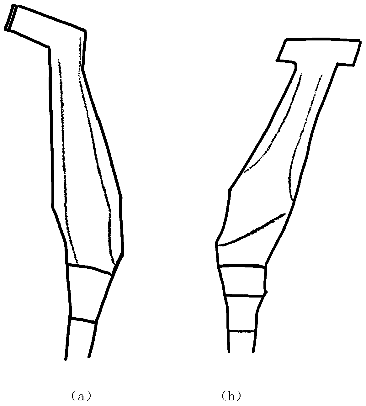
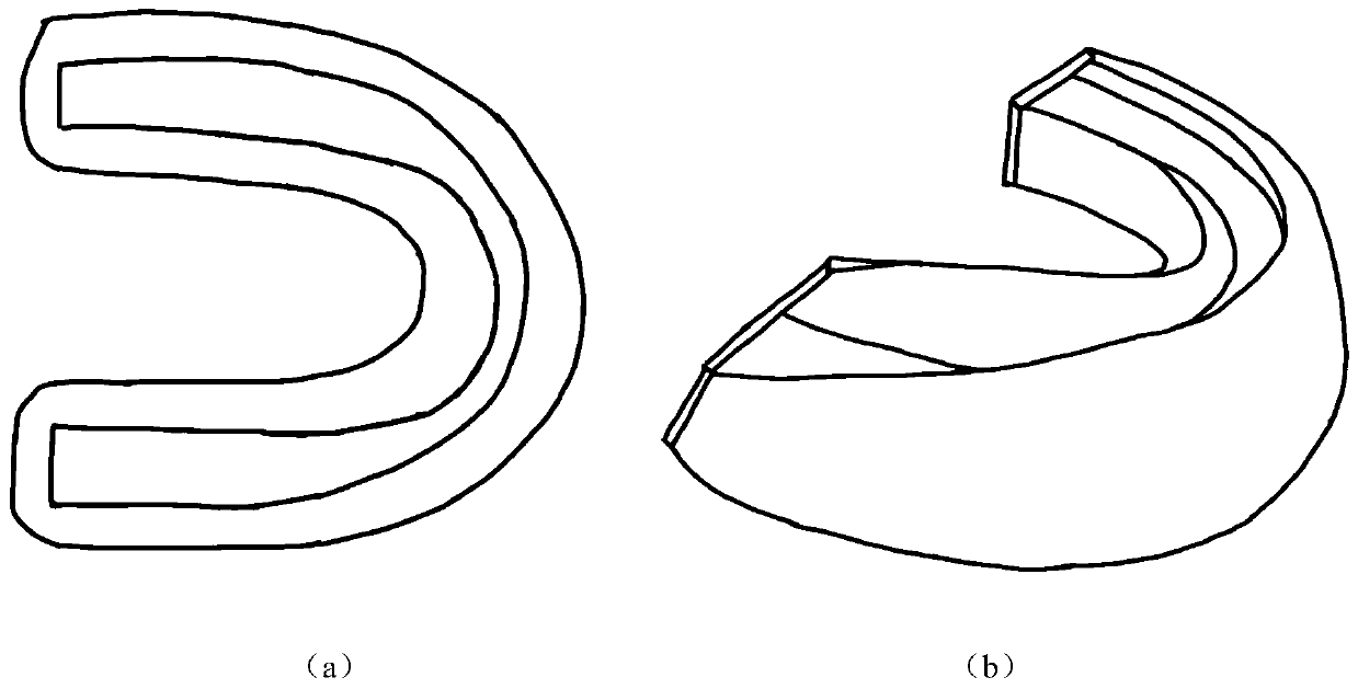
[0030] refer to Figure 1 ~ Figure 3 , an oral cavity scanning device based on high-frequency ultrasound, the device is a single-element ultrasonic probe with a position sensor and a gel brace made of ultrasonic gel or a single-element ultrasonic probe fixed to a manipulator.

[0031] Further, the position sensor includes but not limited to mechanical and electromagnetic position sensors.

[0032] Still further, the surface preparation materials of the gel dental braces include but not limited to ultrasonic-permeable materials such as silica gel, and the filling materials of the gel dental braces include but not limited to ultrasonic gel, deionized water, etc. s material.

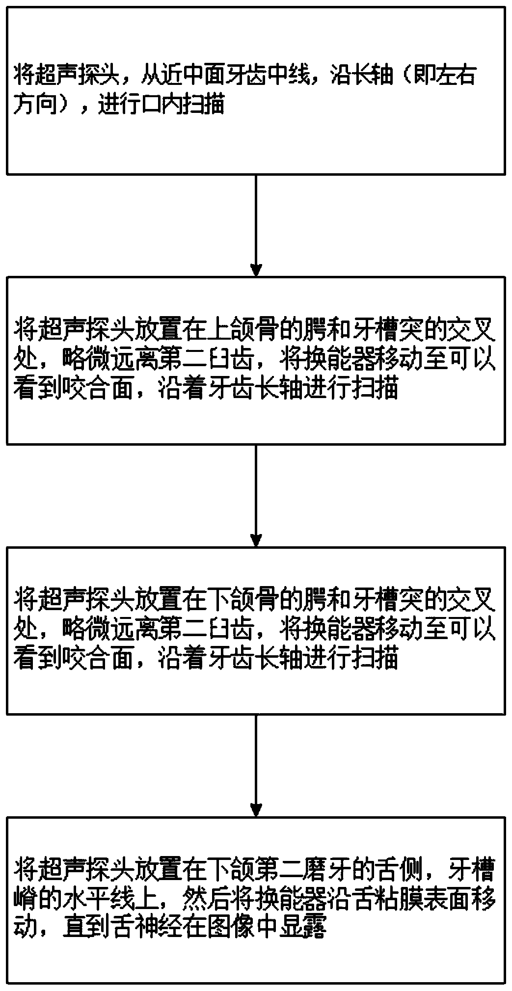
[0033] A method for oral cavity scanning based on high-frequency ultrasound, the method comprising the following steps: 1) using an oral cavity scanning device based on high-frequency ultrasound t...


PUM


Abstract

The invention relates to an oral cavity scanning device and method based on high-frequency ultrasonic. The device is single-vibration-unit ultrasonic probe or a single-vibration-unit ultrasonic probefixed to a mechanical arm, wherein the single-vibration-unit ultrasonic probe is provided with a position sensor and utilizes a gel tooth socket made of ultrasonic gel. The method includes the steps of firstly, using the oral cavity scanning device to scan teeth and periodontal tissue face by face; secondly, using an ultrasonic image segmenting and rebuilding technology to segment and rebuild a target tooth section. The oral cavity scanning device and method has the advantages that tooth and periodontal tissue rebuilding can be achieved in a fast, efficient and zero radiation manner, image acquisition speed and accuracy in oral medicine are increased greatly, and the device and method is high in clinical practical value.

Description

technical field [0001] The invention belongs to the field of medical detection and medical equipment, and relates to an oral cavity scanning device and method. Background technique [0002] In the current clinical practice of stomatology, the methods of obtaining dental conditions mainly rely on making dental models, optical examinations and X-ray examinations. The dental model can visually see the condition of all the crowns, but the production is relatively time-consuming, has many steps, and will be affected by the oral environment. X-rays can diagnose some problems that are not noticeable in appearance, and can accurately see whether there is any abnormality in the condition of teeth. However, X-ray imaging is powerless for some hard tissue loss. Although the radiation dose of modern dental equipment is small, X-rays cannot be used for a long time, especially for patients such as pregnant women. Basically, X-rays are only used in emergency situations. Optical scanning ...

Claims


Application Information

Patent Timeline
Patent Type & Authority Applications(China)
IPC IPC(8): A61B8/12A61B8/00
CPCA61B8/12A61B8/4444A61B8/5223
Inventor 姜娓娓高情毓陈贤挺高飞
Owner ZHEJIANG UNIV OF TECH
Why Eureka
Try Eureka