Abdomen ultrasonic image segmentation method

An ultrasound image and abdominal technology, applied in the field of image processing, can solve the problems of complex segmentation, time-consuming, slow image segmentation, etc., and achieve the effect of increasing the scale and range, speeding up the segmentation speed, and simplifying the process

Pending Publication Date: 2020-02-28
SHANGHAI UNIV OF ENG SCI
View PDF5 Cites 5 Cited by
  • Summary
  • Abstract
  • Description
  • Claims
  • Application Information

AI Technical Summary

Problems solved by technology

Therefore, part of the time is spent in the process of cutting the small images in the training process, and most of the time is spent in the process of cutting the small images and splicing them into the whole image in the test process, that is, there is a problem of complex segmentation and too much time. It leads to slow image segmentation; in addition, the small images after cutting need to be stored for training, which will cause additional waste of computer storage resources

Method used


Image

  • Abdomen ultrasonic image segmentation method
  • Abdomen ultrasonic image segmentation method
  • Abdomen ultrasonic image segmentation method

Examples


Embodiment Construction

[0030] The present invention will be described in detail below in conjunction with the accompanying drawings and specific embodiments.

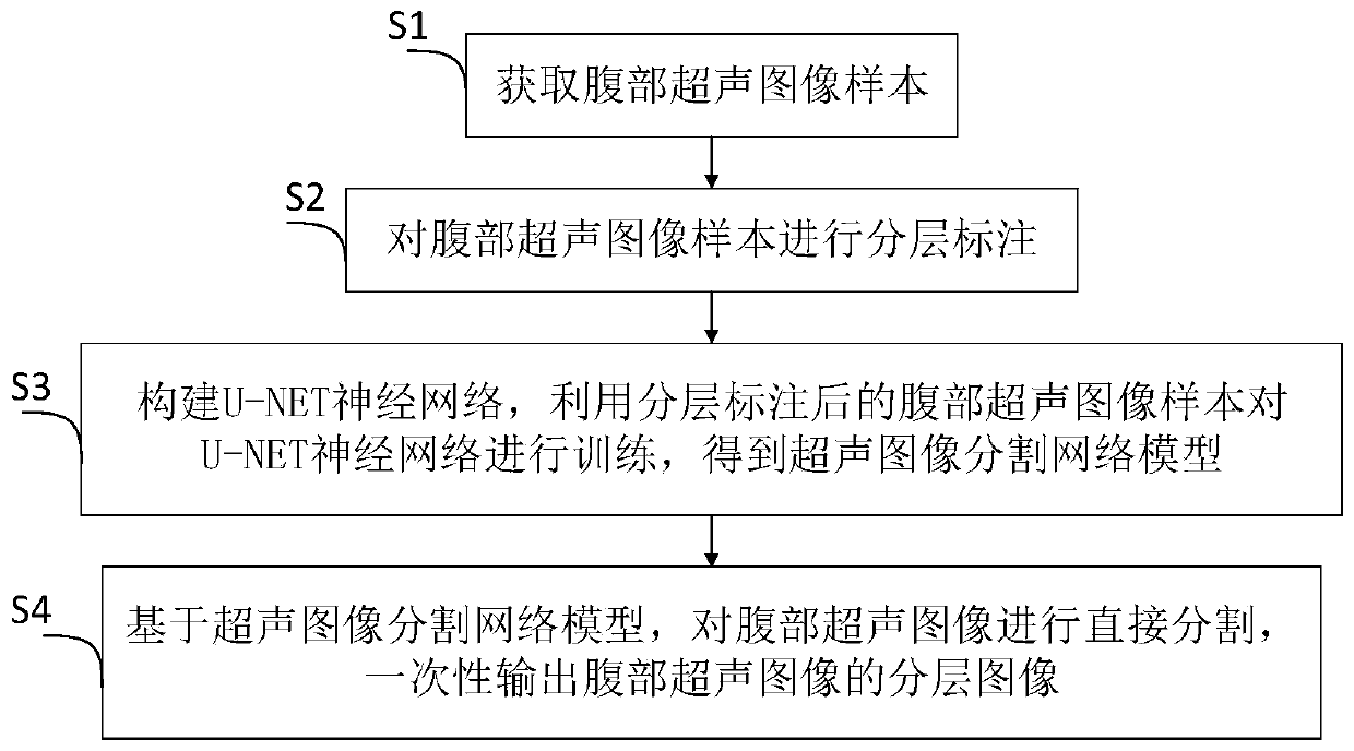
[0031] Such as figure 1 Shown, a kind of abdominal ultrasound image segmentation method comprises the following steps:

[0032] S1. Acquiring abdominal ultrasound image samples;

[0033] S2. Hierarchically labeling the abdominal ultrasound image samples;

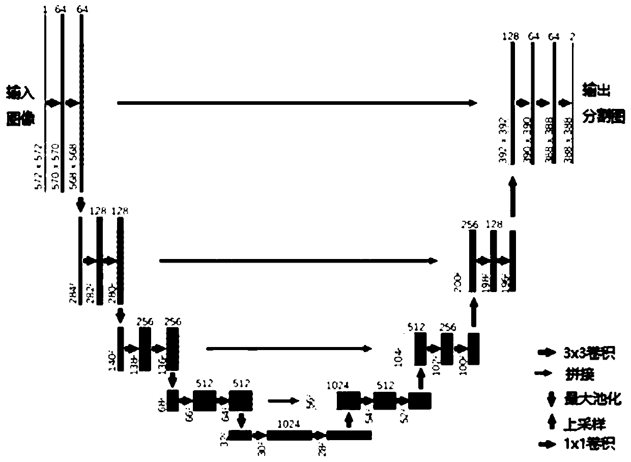
[0034] S3. Construct the U-NET neural network, use the abdominal ultrasound image samples after layered labeling to train the U-NET neural network, and obtain the ultrasound image segmentation network model;

[0035] S4. Based on the ultrasonic image segmentation network model, the abdominal ultrasonic image is directly segmented, and a layered image of the abdominal ultrasonic image is output at one time.

[0036] In practical application, the above method can be realized by the abdominal ultrasound image acquisition module, the segmentation network establishment module and the abdomina...


PUM


Abstract

The invention relates to an abdomen ultrasonic image segmentation method. The method comprises the following steps: S1, acquiring an abdomen ultrasonic image sample; S2, performing layered labeling onthe abdomen ultrasonic image sample; S3, constructing a U-NET neural network, and training the U-NET neural network by using the abdomen ultrasonic image sample subjected to layered labeling to obtain an ultrasonic image segmentation network model; and S4, based on the ultrasonic image segmentation network model, directly segmenting the abdominal ultrasonic image, and outputting a layered image of the abdominal ultrasonic image at one time. Compared with the prior art, the scale quantity and range of training samples are increased through data expansion, the step of cutting small images is avoided, meanwhile, computer storage resources are saved, end-to-end learning is conducted through the full convolutional neural network, the segmentation precision of all layers of the abdomen is higher, the segmentation results of all layers in the abdomen ultrasonic image can be output at a time, and the segmentation speed is effectively increased.

Description

technical field [0001] The invention relates to the technical field of image processing, in particular to a method for segmenting abdominal ultrasound images. Background technique [0002] Image segmentation is the technology and process of dividing the image into several specific and unique regions, and extracting the target of interest. By segmenting the abdominal ultrasound image, the main layers of the abdomen can be automatically distinguished: skin layer, fat Layer, subcutaneous muscle layer and peritoneum, thereby reducing the identification of artificial naked eyes, which is conducive to rapid guided ultrasound puncture. [0003] Since the neural network has the algorithm characteristics of simulating human perception, the segmentation performance is significantly improved compared with other algorithms. At present, the ultrasound image is usually segmented by means of the neural network, but this segmentation method generally adopts the mode of cutting into small im...

Claims


Application Information

Patent Timeline
Patent Type & Authority Applications(China)
IPC IPC(8): G06T7/11G06T5/00
CPCG06T7/11G06T5/002G06T2207/10132G06T2207/20081G06T2207/20084G06T2207/30004
Inventor 方志军顾佳高永彬田方正董九庆
Owner SHANGHAI UNIV OF ENG SCI
Why Eureka
Try Eureka