
Bionic liver organ culture model and application thereof

An organoid and model technology, which is applied in the field of in vitro cell culture technology and drug toxicity screening to achieve the effects of unique structure, enhanced toxicity sensitivity, and enhanced liver specific function and metabolic function.

Pending Publication Date: 2020-04-03
江苏信安佳医疗科技有限公司
View PDF0 Cites 4 Cited by
  • Summary
  • Abstract
  • Description
  • Claims
  • Application Information

AI Technical Summary

Problems solved by technology

However, there is currently no research on the co-culture model of hepatocytes and endothelial cells and its use in drug toxicity screening, especially the co-culture model of three-dimensional hepatocyte spheroids and endothelial cells has not been tried and reported.

Method used


Image

  • Bionic liver organ culture model and application thereof
  • Bionic liver organ culture model and application thereof
  • Bionic liver organ culture model and application thereof

Examples


Embodiment 1

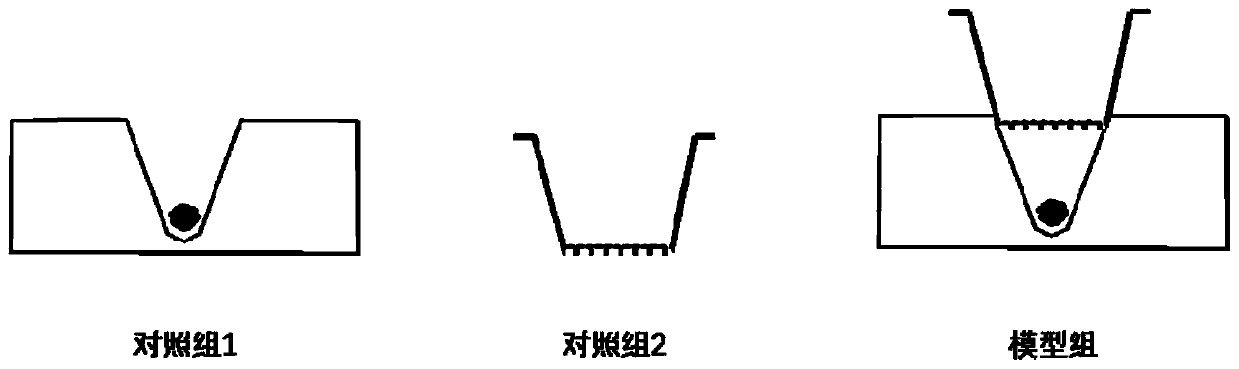
[0044] Example 1: Construction of a three-dimensional hepatocyte spheroid culture model alone (control group 1):

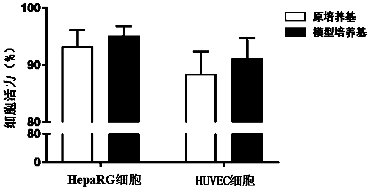
[0045] The hepatocytes were human hepatic progenitor cells HepaRG (purchased from Thermo Fisher), and the HepaRG cells were cultured according to the medium specified in the instructions (purchased from Thermo Fisher). HepaRG cells were digested with trypsin, and the cell viability was detected by trypan blue. The cell viability reached more than 90%, and it was ready for use.

[0046] Pipette the HepaRG cell suspension evenly and add it into the groove device, place it in a 37°C, 5% CO2 incubator for cultivation, and cultivate for 24 hours to form a three-dimensional hepatocyte spheroid culture model alone, and replace the culture every 2 days liquid.

Embodiment 2

[0047] Embodiment 2: Construction of endothelial cell culture model alone (control group 2):

[0048] The endothelial cells are human endothelial cells HUVEC (purchased from ATCC), and the HUVEC cells are cultured according to the medium specified in the instructions (purchased from ATCC). The HUVEC cells were digested with trypsin, and the cell viability was detected by trypan blue, and the cell viability reached more than 90%, and it was ready for use.

[0049] Pipette the HUVEC cell suspension evenly and add it to the tranwell, place it in a 37°C, 5% CO2 incubator for cultivation, and cultivate for 24 hours to form an endothelial cell culture model alone, and replace the culture medium every 2 days.

Embodiment 3

[0050] Example 3: Construction of a bionic liver organoid culture model (model group):

[0051] The hepatocytes were human hepatic progenitor cells HepaRG (purchased from Thermo Fisher), and the HepaRG cells were cultured according to the medium specified in the instructions (purchased from Thermo Fisher). The endothelial cells are human endothelial cells HUVEC (purchased from ATCC), and the HUVEC cells are cultured according to the medium specified in the instructions (purchased from ATCC). HepaRG and HUVEC cells were digested with trypsin, respectively, and the cell viability was detected by trypan blue. The viability of HepaRG and HUVEC cells reached more than 90%, and they were set aside.

[0052] Pipette the HepaRG cell suspension evenly and add it into the groove device, place it in a 37°C, 5% CO2 incubator for cultivation, and cultivate for 24 hours to form a three-dimensional hepatocyte spheroid culture model alone. The HUVEC cell suspension was pipetted evenly and ad...


PUM


Abstract

The invention relates to the fields of cell in-vitro culture technology and drug toxicity screening, in particular to construction of a bionic liver organ culture model and an application of the bionic liver organ culture model in drug toxicity evaluation. The invention provides a construction method of the bionic liver organ culture model, which comprises the following steps: respectively carrying out conventional cell culture and digestion on hepatocytes and endothelial cells to prepare cell suspensions, respectively inoculating the cell suspensions to a groove device and a porous membrane,assembling the porous membrane to the groove device, and culturing in an incubator to prepare the bionic liver organ culture model. The model is unique in structure, has double cells and double targets, can be used for toxicity evaluation of hepatocytes with main targets and toxicity evaluation of non-parenchyma endothelial cells, and provides a new thought and a powerful tool for drug toxicity screening.

Description

technical field [0001] The invention relates to the fields of cell in vitro culture technology and drug toxicity screening, in particular to the construction of a bionic liver organoid culture model and its application for drug toxicity assessment. Background technique [0002] Drug-induced liver injury (DILI) is one of the main reasons why drug development fails and is withdrawn after marketing. Since animal models lack accurate predictability and vigorously implement the 3R principle (ie, replace, reduce, optimize), it is particularly important to establish a reliable in vitro human-derived hepatocyte model and use it for drug hepatotoxicity screening research. However, studies have found that hepatocytes tend to lose their structural characteristics and differentiation ability during routine culture in vitro, mainly because hepatocytes lack the three-dimensional structure and intercellular interactions of the liver in vivo, which leads to the reduction of liver function a...

Claims


Application Information

Patent Timeline
IPC IPC(8): C12N5/071C12Q1/02
CPCC12N5/0671C12N5/0602G01N33/5008G01N33/5064G01N33/5067C12N2533/30
Inventor 罗国安马立东王乙同王义明
Owner 江苏信安佳医疗科技有限公司
Features
  • Generate Ideas
  • Intellectual Property
  • Life Sciences
  • Materials
  • Tech Scout
  • Unparalleled Data Quality
  • Higher Quality Content
  • 60% Fewer Hallucinations
Learn More