
Head and neck lymph node visual display method and device and storage medium

A lymph node, head and neck technology, applied in the field of visual display of head and neck lymph nodes, can solve the problems of changing tissues, unable to visually see different areas, differences in the spread of different cancer types, and unable to accurately display or explain the development process of the disease. , to reduce the occupancy rate, facilitate observation and learning, and enhance the visualization effect.

Active Publication Date: 2020-04-14
PERCEPTION VISION MEDICAL TECH CO LTD +1
View PDF6 Cites 0 Cited by
  • Summary
  • Abstract
  • Description
  • Claims
  • Application Information

AI Technical Summary

Problems solved by technology

[0003] However, the above method is not only difficult to make students fully grasp all types and stages of head and neck lymph node lesions; moreover, for the first anatomy or surgical operation, head and neck lymph nodes are the most complex in structure, and it is necessary to have Physicians with certain surgical skills can fully present the diseased tissue. During the dissection process, the position of the tissue may be changed, affecting the judgment of the axis and plane. Students rarely have the opportunity to do it themselves, and cannot achieve the purpose of directly observing its structure; for The second method of observing human tissue samples can only study local tissues, and cannot intuitively see the difference in the spread of different regions and different types of cancer; for the third method of observing medical imaging, although it can be seen very well The state of the original tissue in the human body can see the difference in the spread of different cancer types, but this data mostly comes from clinical data, and the data of different stages may be inconsistent or lacking (for example: oropharyngeal cancer, the first-stage image in A The second phase of the image was taken in hospital B, the third phase did not undergo re-examination, and the fourth phase of the image was taken in A hospital), which cannot accurately and systematically show or explain the development of the disease process to students

Method used


Image

  • Head and neck lymph node visual display method and device and storage medium
  • Head and neck lymph node visual display method and device and storage medium
  • Head and neck lymph node visual display method and device and storage medium

Examples


Embodiment 1

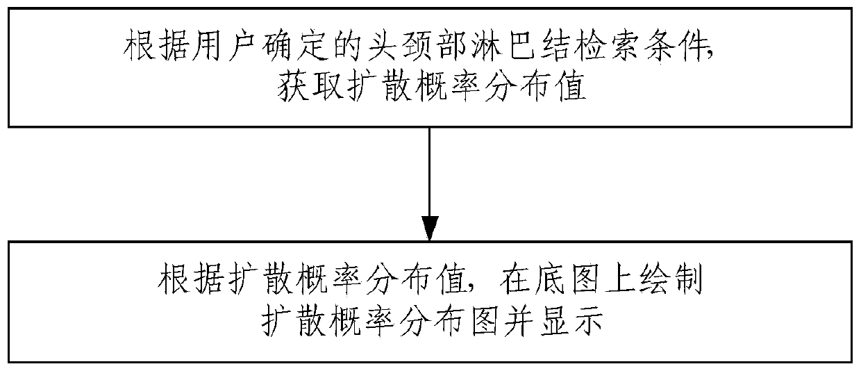
[0027] A visual display method for head and neck lymph nodes provided by an embodiment of the present invention, such as figure 1 As shown, the method includes:

[0028] According to the head and neck lymph node retrieval conditions determined by the user, the diffusion probability distribution value is obtained;

[0029] According to the diffusion probability distribution value, the diffusion probability distribution map is drawn on the base map and displayed.

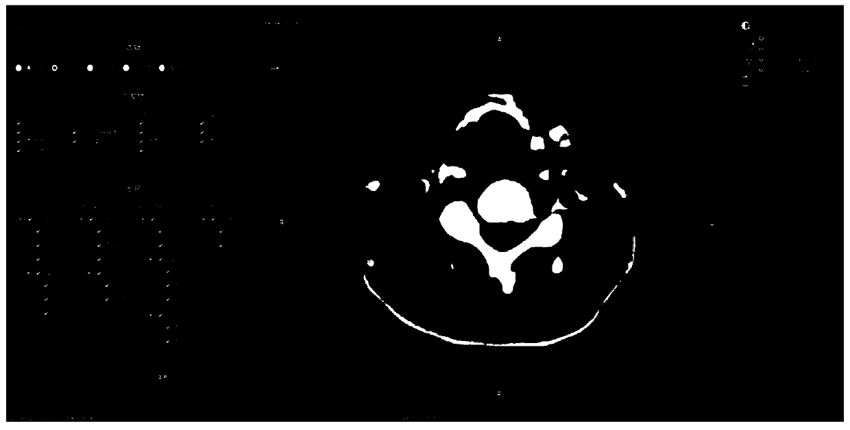
[0030] Specifically, the retrieval conditions include but are not limited to: cancer type, primary tumor and stage. For better illustration, the above method is applied to a computer system, the operation interface of which is as follows figure 2 As shown, including the search area on the left and the drawing area on the right. The method for the user to determine the head and neck lymph node retrieval condition is: the user selects through the linkage screening logic provided in the retrieval area on the operatio...

Embodiment 2

[0045] An embodiment of the present invention provides a visual display device for head and neck lymph nodes, including a retrieval module, used for users to input head and neck lymph node retrieval conditions, and obtain diffusion probability distribution values; Drawing and displaying a diffusion probability distribution map; and a preset data storage module, used for storing preset diffusion probability distribution values. Preferably, the device uses the method described in Example 1.

[0046] Specifically, the operation interface of the device is as follows figure 2 As shown, including the search area on the left and the drawing area on the right. The search area adopts a linkage screening method for users to input head and neck lymph node search conditions; the drawing area provides real-time and visualized images of head and neck lymph nodes according to the search conditions determined on the left.

[0047] Users can conveniently input and confirm the search conditi...

Embodiment 3

[0049] The embodiment of the present invention also provides an electronic device, such as Figure 7 As shown, the electronic device may include: a processor (processor) 301, a communication interface (Communications Interface) 302, a memory (memory) 303, and a communication bus 304, wherein the processor 301, the communication interface 302, and the memory 303 pass through the communication bus 304 Complete mutual communication. The processor 301 can invoke a computer program stored in the memory 303 and runnable on the processor 301 to execute the method provided by the above-mentioned embodiment, for example, including: acquiring the diffusion probability distribution value according to the head and neck lymph node retrieval condition determined by the user ;According to the diffusion probability distribution value, draw the diffusion probability distribution map on the base map and display it.

[0050] In addition, the above logic instructions in the memory 303 may be imp...


PUM


Abstract

The embodiment of the invention provides a head and neck lymph node visual display method and device and a storage medium, and the method comprises the steps: obtaining a diffusion probability distribution value according to a head and neck lymph node retrieval condition determined by a user; and drawing a diffusion probability distribution map on the base map according to the diffusion probability distribution value and displaying the diffusion probability distribution map. According to the embodiment of the invention, the lymph node distribution form and diffusion difference can be displayedin real time according to the head and neck lymph node retrieval condition determined by a user; by displaying partition boundary lines, a focus change trend is convenient to observe; by displaying equal probability lines, teachers and students can conveniently and accurately locate the diffusion probability distribution range; a user can quickly and continuously switch medical images of different cancer types, primary lesions and stages only by modifying retrieval conditions, and different lesions and extended differences are accurately presented; and data calling and drawing are performed based on an equal probability line and an equal probability plane, so that the occupancy rate of hardware resources is reduced, and the visualization effect is enhanced.

Description

technical field [0001] The invention relates to the technical field of medical teaching, in particular to a visual display method, device and storage medium of head and neck lymph nodes. Background technique [0002] During the teaching and training process, imaging doctors can accurately and efficiently grasp the clinical identification and diagnosis of the disease by visually observing the distribution and diffusion of head and neck lymph nodes under different cancer types, different primary lesions, and different stages. Diagnostic skills, so as to quickly achieve the goal of training and teaching. The existing teaching and training are mainly carried out through the following means: first, through dissection, surgical operation, etc., to directly observe the lesion tissue structure, or by watching the anatomy and operation process recorded by video; The third is to observe medical images such as CT, PET, etc., to observe, study, explain and teach the images. [0003] H...

Claims


Application Information

Patent Timeline
Patent Type & Authority Applications(China)
IPC IPC(8): G16H30/40
CPCG16H30/40Y02A90/10
Inventor 李浩鹏矫承洋陈桂铭吴健伟吕丽云魏军
Owner PERCEPTION VISION MEDICAL TECH CO LTD
Learn More