CT image lung lobe method and system

A CT image and lung technology, applied in the fields of medical image processing and artificial intelligence, can solve problems such as differences in lobulation results, long time-consuming lung lobes, and difficulty in recording and displaying, and achieve the effect of improving processing efficiency

Inactive Publication Date: 2020-06-09
北京精诊医疗科技有限公司
View PDF4 Cites 9 Cited by
  • Summary
  • Abstract
  • Description
  • Claims
  • Application Information

AI Technical Summary

Problems solved by technology

[0005] In order to solve the existing problems of manually distinguishing lung lobes, such as long time-consuming, differences in lobulation results, and difficulty in recording and displaying, the present invention provides a method for lung lobes in CT images, including:

Method used


Image

  • CT image lung lobe method and system
  • CT image lung lobe method and system
  • CT image lung lobe method and system

Examples


Embodiment Construction

[0043] The technical solutions of the present invention will be further described below in conjunction with the accompanying drawings and embodiments.

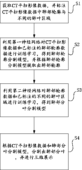
[0044] see figure 1 The CT image lung lobulation method provided by the embodiment of the present invention specifically includes the following steps:

[0045] Step S1. Obtain CT plain scan image data, and label the lung contour and different lung lobe regions in the CT plain scan image data.

[0046] In this embodiment, 1000 cases of CT plain scan image data are collected, including 400 cases of healthy CT plain scan image data, 300 cases of pneumonia CT plain scan image data and 300 cases of pulmonary nodule CT plain scan image data, of which 800 cases are selected as training data , 200 cases were used as test data, and the lung contour and different lung lobe regions in the CT plain scan image data were marked. The specific process is as follows:

[0047] Step S101 , acquiring plain CT scan image data, and cleaning the p...


PUM


Abstract

The invention discloses a CT image lung lobe method and system, and belongs to the technical field of medical image processing and artificial intelligence. The method comprises the following steps: marking a lung contour and a lung lobe area in CT horizontal scanning image data; according to the CT horizontal scanning image data and the lung contour data, performing training learning by using a first neural network to obtain a lung contour segmentation model; extracting the lung contour according to the lung contour segmentation model, and performing training learning on the lung contour dataand the lung lobe area by using a second neural network to obtain a lung partial lobe segmentation model; and according to the CT horizontal scanning image data and the lung part leaf segmentation model, segmenting lung part leaves, and performing three-dimensional display. The system comprises a labeling module, a first training module, a second training module and a segmentation display module.According to the CT image lung lobe method and system, the processing efficiency of CT image data is improved, the lung area and the lung part leaves can be quickly segmented, and the segmented lung part leaves are displayed in a three-dimensional mode.

Description

technical field [0001] The invention relates to the technical fields of medical image processing and artificial intelligence, in particular to a CT image lung lobulation method and system. Background technique [0002] The human lungs are located in the thoracic cavity, suspended on both sides of the mediastinum by the lung roots and pulmonary ligaments. All organs of the human body cannot work without oxygen, and the lungs are the only organ that transports oxygen and discharges carbon dioxide. Through breathing, the alveoli will be in contact with the atmosphere at any time and perform gas exchange, inhaling oxygen and exhaling carbon dioxide. The human body has two lungs, one on the left and one on the left, and the surface of the lungs is covered with the pleura and the visceral layer of the septum. The left lung is long and narrow, and is divided into upper and lower lobes by oblique fissures; the right lung is wide and short, and is divided into upper, middle, and low...

Claims


Application Information

Patent Timeline
Patent Type & Authority Applications(China)
IPC IPC(8): G06T7/11G06T17/00
CPCG06T7/11G06T17/00G06T2207/10081G06T2207/20081G06T2207/20084G06T2207/20132G06T2207/30061
Inventor 王博赵威代笃伟侯雪雪徐正清金烁申建虎张伟金洪波靳博方潘承燕
Owner 北京精诊医疗科技有限公司
Why Eureka
Try Eureka