Magnetic resonance angiography method, device and equipment

A technology of vascular imaging and magnetic resonance, applied in medical science, sensors, diagnostic recording/measurement, etc., can solve the problems of increasing the number of scans and reducing the efficiency of MRA imaging, and achieve the effect of improving efficiency

Active Publication Date: 2020-06-12
SHANGHAI UNITED IMAGING HEALTHCARE
View PDF6 Cites 9 Cited by
  • Summary
  • Abstract
  • Description
  • Claims
  • Application Information

AI Technical Summary

Problems solved by technology

[0003]In order to obtain more accurate blood flow velocity information, it is usually necessary to perform multiple repeated magnetic resonance scans on the measured part based on one acquisition method, but such a technical method Reduced MRA imaging efficiency due to increased number of scans

Method used


Image

  • Magnetic resonance angiography method, device and equipment
  • Magnetic resonance angiography method, device and equipment
  • Magnetic resonance angiography method, device and equipment

Examples


Embodiment 1

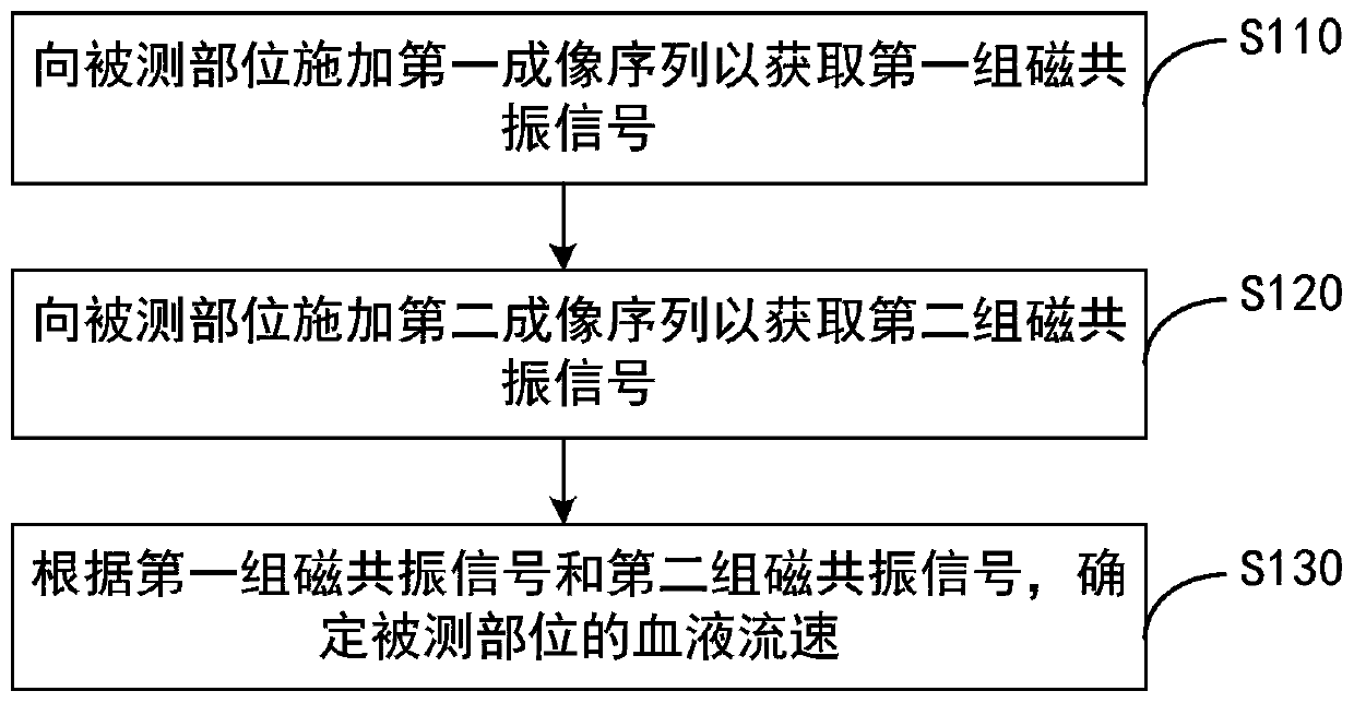
[0039] figure 1 It is a flow chart of a magnetic resonance angiography method provided in Embodiment 1 of the present invention. This embodiment is applicable to the situation where magnetic resonance equipment is used for blood vessel imaging. The method can be performed by a magnetic resonance angiography device, which can Realized by means of software and / or hardware, the device can be configured in magnetic resonance equipment. Specifically include the following steps:

[0040] S110. Apply a first imaging sequence to the measured part to acquire a first set of magnetic resonance signals, wherein the measured part includes blood flow, the first imaging sequence includes a first inversion recovery radio frequency pulse and a first gradient pulse, and the first gradient The pulse is used to suppress the phase information of the blood flow.

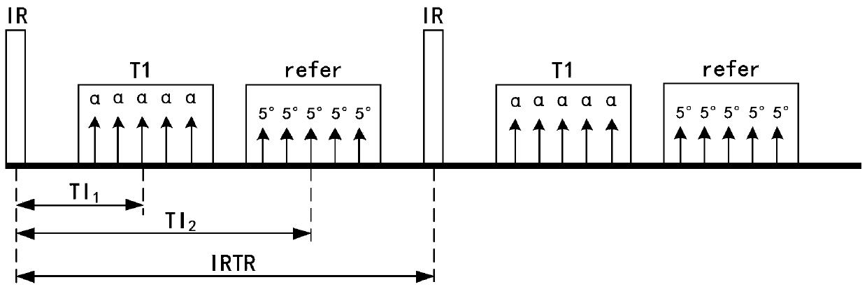
[0041] Simultaneous Non-Contrast Angiography and Intraplaque Hemorrhage Imaging (SNAP) sequence is a phase-sensitive inversion recover...

Embodiment 2

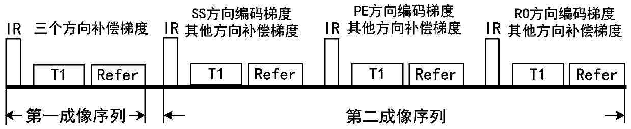
[0090] Figure 6 It is a flow chart of a magnetic resonance angiography method provided in Embodiment 2 of the present invention, and the technical solution of this embodiment is further refined on the basis of the foregoing embodiments. Optionally, the method further includes: according to the magnetic resonance signal acquired based on the T1 acquisition pulse and the magnetic resonance signal acquired based on the refer acquisition pulse in the first set of magnetic resonance signals, determining the vascular morphology image of the measured part , wherein the vascular morphology image includes at least one of intraplaque hemorrhage image, vessel wall image, vessel image and real part image.

[0091] The specific implementation steps of this embodiment include:

[0092] S210. Apply a first imaging sequence to the measured part to acquire a first set of magnetic resonance signals, wherein the measured part includes blood flow, the first imaging sequence includes a first inv...

Embodiment 3

[0105] Figure 8 It is a schematic diagram of a magnetic resonance angiography apparatus provided in Embodiment 3 of the present invention. This embodiment is applicable to the case of using magnetic resonance equipment to perform blood vessel imaging, and the apparatus can be implemented in software and / or hardware, and the apparatus can be configured in the magnetic resonance equipment. The magnetic resonance angiography apparatus includes: a first group of magnetic resonance signal acquisition module 310 , a second group of magnetic resonance signal acquisition module 320 and a blood flow velocity determination module 330 .

[0106] Wherein, the first group of magnetic resonance signal acquisition module 310 is configured to apply a first imaging sequence to the measured part to obtain the first group of magnetic resonance signals, wherein the measured part includes blood flow, and the first imaging sequence includes a first inversion recovering the radio frequency pulse a...


PUM


Abstract

The embodiment of the invention discloses a magnetic resonance angiography method, a device and equipment. The method comprises the following steps that: a first imaging sequence is applied to a measured part to obtain a first set of magnetic resonance signals, wherein the measured part comprises blood flow, the first imaging sequence comprises a first inversion recovery radio frequency pulse anda first gradient pulse, and the first gradient pulse is used for inhibiting phase information of the blood flow; a second imaging sequence is applied to the measured part to obtain a second set of magnetic resonance signals, wherein the second imaging sequence comprises a second inversion recovery radio frequency pulse and a second gradient pulse, and the second gradient pulse is used for obtaining phase information of the blood flow; and the blood flow velocity of the measured part is determined according to the first set of magnetic resonance signals and the second set of magnetic resonancesignals. According to the embodiment of the invention, the imaging sequence is improved, so that the problem that magnetic resonance scanning needs to be repeated for multiple times is solved, and themagnetic resonance angiography efficiency is improved.

Description

technical field [0001] Embodiments of the present invention relate to the technical field of magnetic resonance imaging, and in particular, to a magnetic resonance angiography method, device, and equipment. Background technique [0002] Magnetic resonance imaging (Magnetic Resonance Imaging) is a method of medical examination and diagnosis that uses hydrogen nuclei in the human body to resonate in a strong magnetic field to generate images. Magnetic resonance angiography (MR Angiography, hereinafter referred to as MRA imaging) is a non-invasive angiography method that does not require intubation and contrast agents. Flow Mechanics Information. [0003] In order to obtain more accurate blood flow velocity information, it is usually necessary to perform multiple repeated magnetic resonance scans on the measured site based on an acquisition method, but such a technical method reduces the efficiency of MRA imaging due to the increased number of scans. Contents of the inventio...

Claims


Application Information

Patent Timeline
Patent Type & Authority Applications(China)
IPC IPC(8): A61B5/0285A61B5/055
CPCA61B5/0263A61B5/0285A61B5/055
Inventor 覃浩东贺强
Owner SHANGHAI UNITED IMAGING HEALTHCARE
Why Eureka
Try Eureka