Visual tracheal intubation kit for small animals

A technology for tracheal intubation and small animals, applied in the medical field, can solve problems such as brain neuron damage, airway tissue fragility, affecting treatment and outcome, etc., to achieve enlarged oral cavity, high intubation success rate, and reduce animal damage effect

Active Publication Date: 2020-06-16
南京市江宁医院
View PDF5 Cites 0 Cited by
  • Summary
  • Abstract
  • Description
  • Claims
  • Application Information

AI Technical Summary

Problems solved by technology

If an unobstructed airway for oxygen supply cannot be established within the golden time window (usually 4-7 minutes) after apnea, brain neurons will be irreversibly damaged, which will greatly affect subsequent treatment and outcome
In the past, there was no complete set of finished equipment for small animal tracheal intubation. Each laboratory designed the intubation according to its own conditions and environmental equipment. Intubation can be performed under direct vision under the illumination of a lamp, under the illumination of a flashlight by an assistant, percutaneous light irradiation in a dark room environment, etc. However, the mouth of small animals is small, and the diameter of the airway under direct vision is only a few millimeters. It is very difficult to intubate directly with the human eye, and the airway tissue is fragile, and the damage caused by blind detection is large, and it is easy to slip over the real airway damage to a part similar to the human "piriform sinus", resulting in intubation failure

Method used


Image

  • Visual tracheal intubation kit for small animals
  • Visual tracheal intubation kit for small animals
  • Visual tracheal intubation kit for small animals

Examples


Embodiment

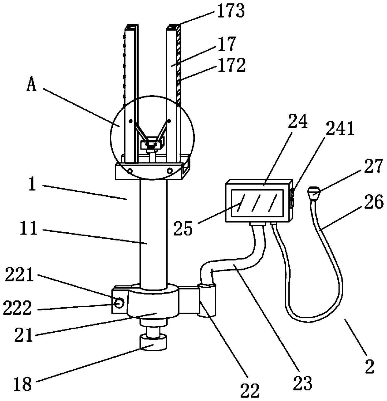
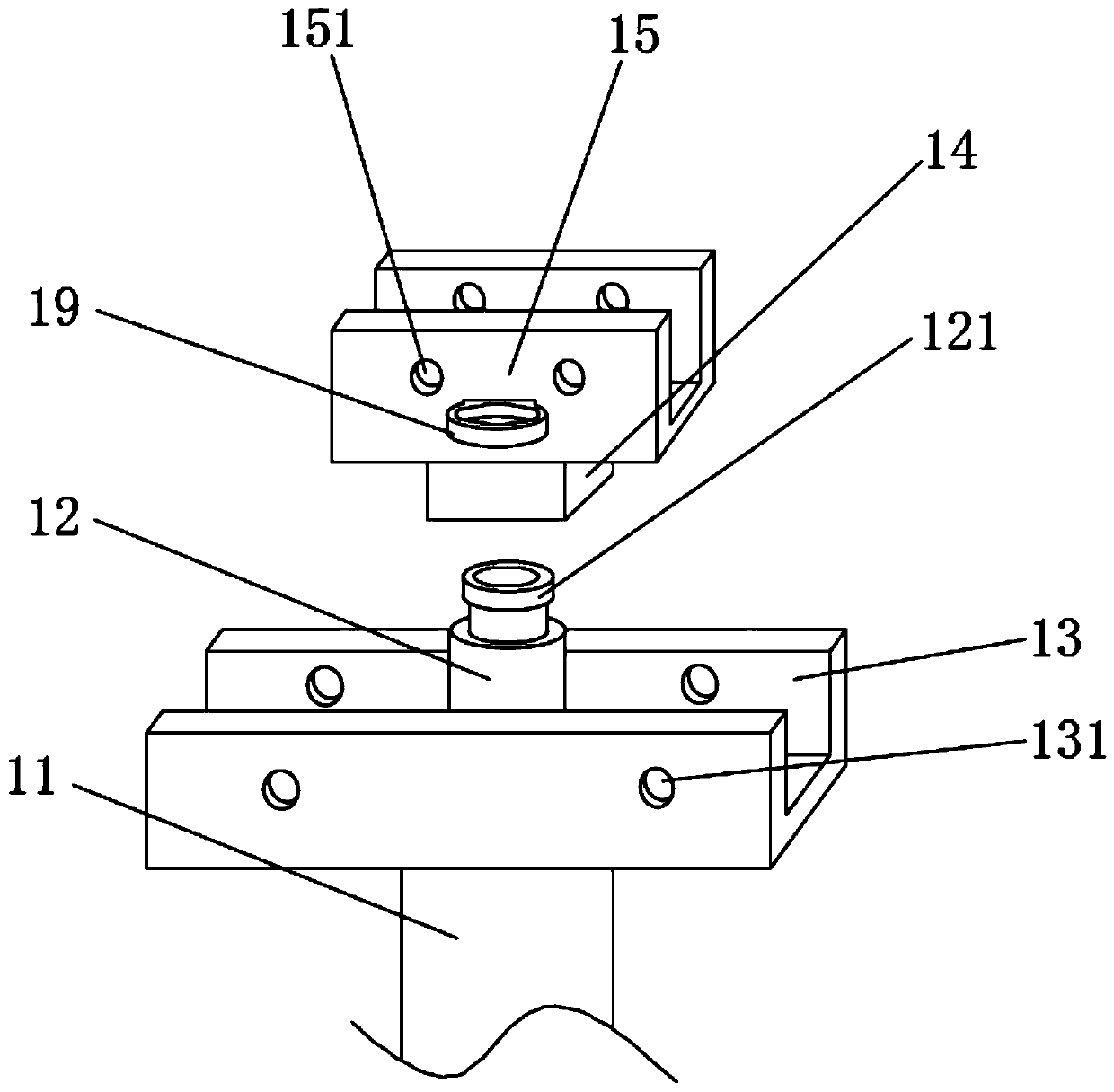
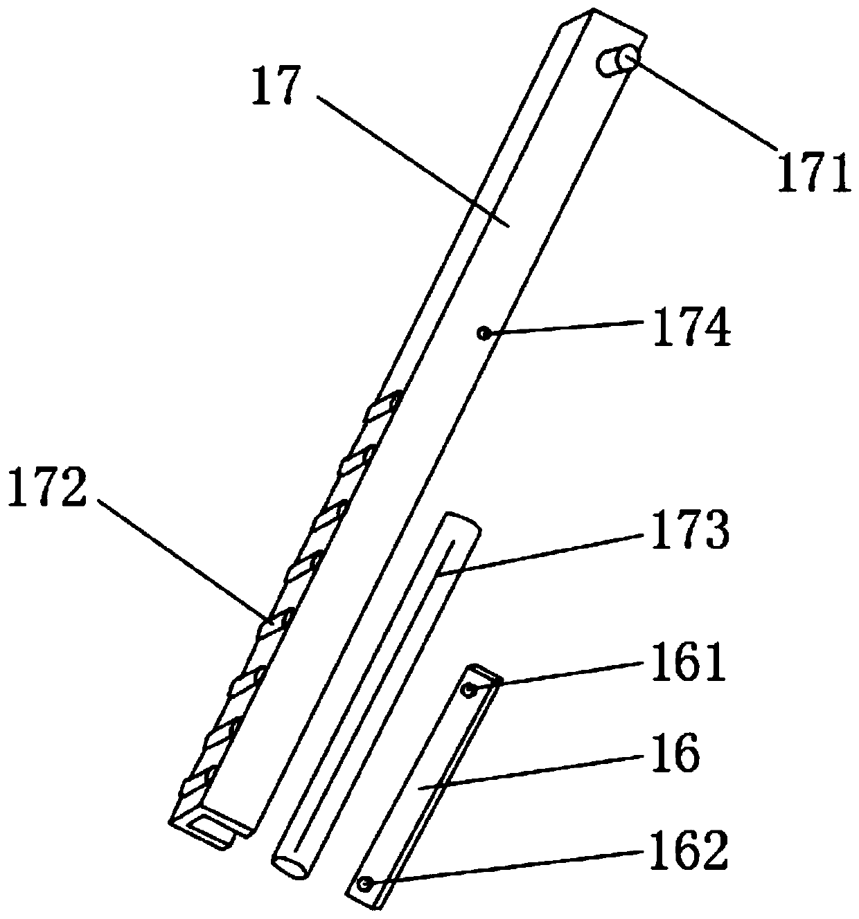
[0023] Example: such as Figure 1-6 As shown, a small animal visual tracheal intubation kit of the present invention includes an oral spreader 1 and a peeping system 2, and the oral spreader 1 includes a hollow handle 11, a hollow screw 12, a No. 1 chuck 13, and a connecting block 14 , No. 2 chuck 15, angle adjusting rod 16 and channel steel 17, the hollow handle 11 is provided with a hollow screw 12, the lower end of the hollow screw 12 is fixedly connected with a knob 18, and the upper end of the hollow screw 12 is fixedly connected with a rotating head 121, The upper end of the hollow handle 11 is fixedly connected with a No. 1 chuck 13, and two No. 1 insertion grooves 131 are respectively opened on both sides of the No. 1 chuck 13. The No. 2 chuck 15 is located above the No. 1 chuck 13. The No. 2 chuck Both sides of No. 15 are provided with two No. 2 embedding grooves 151, the outer wall of No. 2 chuck 15 is fixedly connected with guide ring 19, and the lower side of No. 2...


PUM


Abstract

The invention discloses a visual trachea intubation kit for small animals. The visual trachea intubation kit for small animals comprises an oral cavity dilator and a peeping system, the oral cavity dilator comprises a hollow handle, a hollow screw rod, a first chuck, a connecting block, a second chuck, an angle adjusting rod and steel channels; the hollow screw rod is sleeved in the hollow handle;the lower end of the hollow screw rod is fixedly connected with a knob; the upper end of the hollow screw rod is fixedly connected with a rotating head; the upper end of the hollow handle is fixedlyconnected with the first chuck; two first embedding grooves are formed in the two sides of the first chuck correspondingly; the second chuck is located above the first chuck; two second embedding grooves are formed in each of the two sides of the second chuck; the visual trachea intubation kit is compact in structure and high in intubation success rate, during specific use, the included angle between the steel channels is adjustable, a plurality of convex strips can are adopted for clamping the teeth of a small animal, a supporting portion is located at the teeth of the small animal, the oralcavity of the small animal cannot be damaged, a magnifying lens plays a role in magnifying the view of the oral cavity, an airway can be found easily, and intubation is facilitated.

Description

technical field [0001] The invention relates to a visual trachea intubation kit, in particular to a small animal visual tracheal intubation kit, which belongs to the medical field. Background technique [0002] The most simple and effective first aid measure in case of cardiac arrest is cardiopulmonary resuscitation. Keeping the airway open is an essential step in cardiopulmonary resuscitation, and only an unobstructed airway can guarantee subsequent oxygen therapy. In many animal experiments, it is necessary to simulate clinical disease models. Whether it is simulated emergency operations or surgical treatment after anesthesia, tracheal intubation and ventilator-assisted oxygen therapy are inseparable. Because small animals (this patent is mainly aimed at rodents) have narrow mouths, many secretions after anesthesia, and long tongues, there is very little room for experimenters to operate. If an unobstructed airway for oxygen supply cannot be established within the golden...

Claims


Application Information

Patent Timeline
Patent Type & Authority Applications(China)
IPC IPC(8): A61D7/00A61D15/00A61M16/04A61B1/24A61B1/06A61B1/04
CPCA61B1/04A61B1/0684A61B1/24A61B2503/40A61D7/00A61D15/00A61M16/04A61M2250/00
Inventor 李茜李娟龙云刘文涛段满林
Owner 南京市江宁医院
Why Eureka
Try Eureka