
Magnetic resonance diffusion tensor brain image analysis method and system for newborns

A diffusion tensor and analysis method technology, applied in image analysis, image generation, medical image and other directions, can solve the problem of not using neonatal DTI image analysis method, and achieve clinical and physiological significance.

Pending Publication Date: 2020-06-19
南京慧脑云计算有限公司
View PDF4 Cites 2 Cited by
  • Summary
  • Abstract
  • Description
  • Claims
  • Application Information

AI Technical Summary

Problems solved by technology

According to relevant technical literature, currently up to 74% of newborn brain studies do not use neonatal special magnetic resonance scanning hardware equipment, and 76% of newborn brain imaging studies do not use special neonatal DTI image analysis methods

Method used


Image

  • Magnetic resonance diffusion tensor brain image analysis method and system for newborns
  • Magnetic resonance diffusion tensor brain image analysis method and system for newborns
  • Magnetic resonance diffusion tensor brain image analysis method and system for newborns

Examples


Embodiment Construction

[0059] The present invention will be described in further detail below with reference to the accompanying drawings and the embodiments of the present invention.

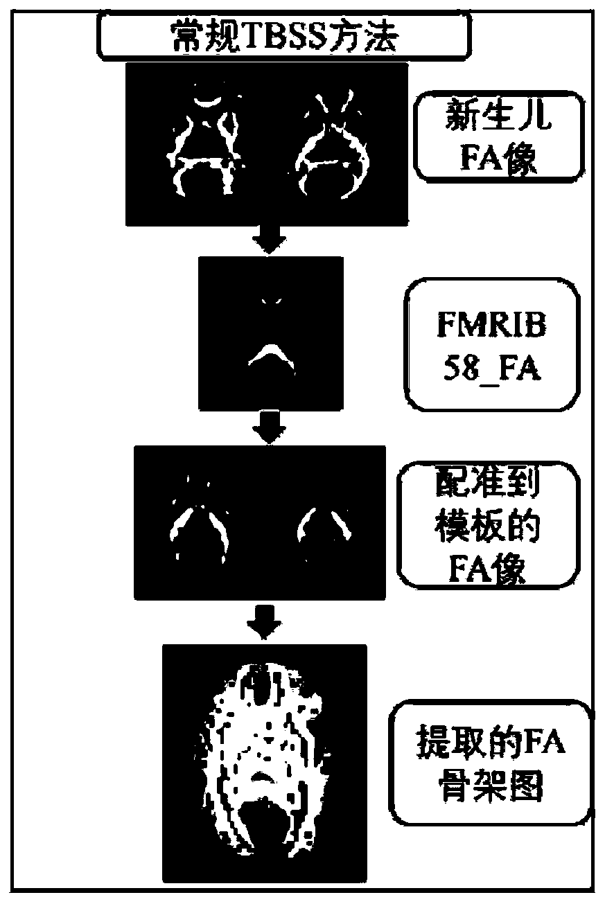
[0060] figure 1 Schematic diagram of the existing process for extracting anisotropy fraction (FA)-based skeletons using conventional spatial-based statistics (TBSS) methods;

[0061] like figure 1 As shown, the process of extracting a skeleton based on anisotropy fraction (FA) using the conventional spatial statistics (TBSS) method mainly includes the following steps:

[0062] The step of preprocessing the neonatal FA image; for the neonatal FMRIB58_FA template, by adopting the algorithm based on spatial statistics (TBSS), the diffusion tensor parameter image of the subject is registered to the template through linear and nonlinear transformation Standard space; Finally, by extracting an anisotropy fraction (FA)-based skeleton map, the problem of mixed brain white matter and gray matter images encountered in conven...


PUM


Abstract

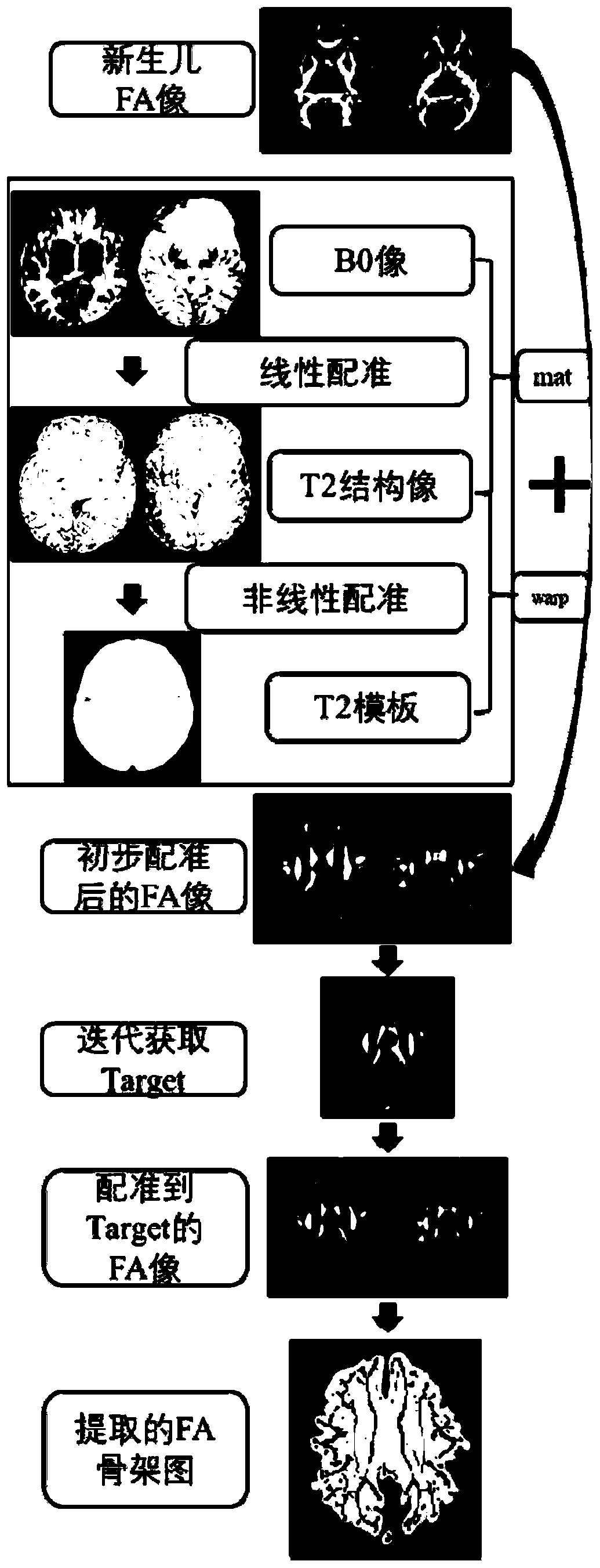
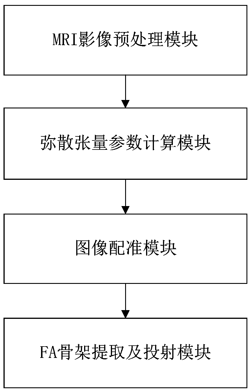
The invention discloses a magnetic resonance diffusion tensor brain image analysis method and system for newborns, and belongs to the field of medical image processing. The method comprises the following steps: A, preprocessing a newborn diffusion tensor imaging DTI image; b, dispersion tensor parameter calculation: fitting the intrinsic value of the dispersion tensor through a DTI tensor model, and calculating a dispersion parameter graph based on an anisotropy fraction FA, an average diffusion coefficient MD, an axial diffusion coefficient AD and a radial diffusion coefficient RD; c, performing image registration; and D, extracting a skeleton based on the anisotropy fraction FA, and projecting the skeleton based on the anisotropy fraction FA, the average diffusion coefficient MD, the axial diffusion coefficient AD and the radial diffusion coefficient RD of the testee onto the skeleton. According to the method, the newborn DTI image is subjected to preprocessing, diffusion tensor parameter calculation, image registration and FA skeleton extraction, and finally the brain image with clear brain white matter and brain gray matter signals is obtained.

Description

technical field [0001] The invention relates to a medical image and a magnetic resonance image (MRI) image processing technology, in particular to a newborn magnetic resonance diffusion tensor brain image analysis method and system thereof. Background technique [0002] Magnetic resonance imaging (MRI) diffusion tensor imaging (Diffusion Tensor Imaging, DTI), clinically also known as diffusion tensor imaging. The principle of DTI is to indirectly observe the microstructural properties of brain white matter tissue by measuring the degree and direction of water molecule diffusion in the tissue. DTI imaging technology is currently the only technology that can display the direction of nerve fiber bundles in the white matter of the brain in a non-destructive way in vivo, so it is widely used in clinical and human brain science research. [0003] Currently the most commonly used and most reliable DTI analysis technique is based on the spatial statistics (Tract-Based SpatialStatis...

Claims


Application Information

Patent Timeline
Patent Type & Authority Applications(China)
IPC IPC(8): G06T7/00G06T7/38G06T11/00G06T3/00G16H30/20
CPCG06T7/0012G06T7/38G06T11/008G16H30/20G06T2207/10088G06T2207/30016G06T2210/41G06T3/04
Inventor 徐明泽廖攀
Owner 南京慧脑云计算有限公司
Learn More