
Chip device for in-situ tissue dyeing and decoloring and use method

A tissue slicing and tissue technology, applied in the field of medical inspection and analysis, can solve the problems of low efficiency, cell surface damage, long time consumption, etc., and achieve the effects of avoiding impurity pollution, short time consumption, and simple operation.

Pending Publication Date: 2022-02-25
NANTONG UNIVERSITY
View PDF0 Cites 1 Cited by
  • Summary
  • Abstract
  • Description
  • Claims
  • Application Information

AI Technical Summary

Problems solved by technology

[0005] Existing tissue decolorization techniques, including photobleaching, acid-base solution elution, and oxidation, all have shortcomings such as low efficiency, long time-consuming, and cell surface damage in the process of tissue decolorization.

Method used


Image

  • Chip device for in-situ tissue dyeing and decoloring and use method
  • Chip device for in-situ tissue dyeing and decoloring and use method
  • Chip device for in-situ tissue dyeing and decoloring and use method

Examples


Embodiment Construction

[0039] Embodiments of the invention are described in detail below, examples of which are illustrated in the accompanying drawings. The embodiments described below by referring to the figures are exemplary only for explaining the present invention and should not be construed as limiting the present invention.

[0040] The present invention adopts the following technical solutions for solving the problems of the technologies described above:

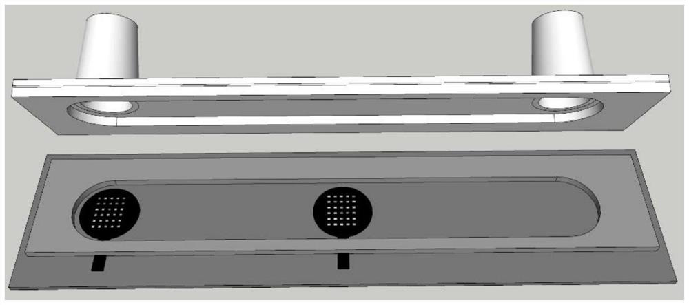
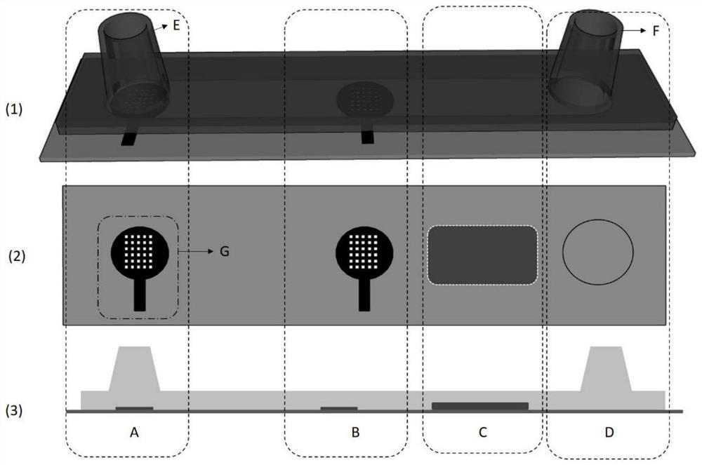
[0041] A chip device for in situ tissue staining and decolorization, such as figure 1 and figure 2 As shown, it includes a three-layer structure that is stacked together and sealed with each other. From top to bottom, it is a top cover, a base plate, and a bottom plate; the materials of the top cover, base plate, and bottom plate are optically transparent and non-biotoxic. Plastic materials including PMMA, polycarbonate, cycloolefin copolymer, or cycloolefin polymer. The thickness of the top cover is 0.5-2.5 mm. The thickness of the s...


PUM

PropertyMeasurementUnit
thicknessaaaaaaaaaa
thicknessaaaaaaaaaa
thicknessaaaaaaaaaa

Abstract

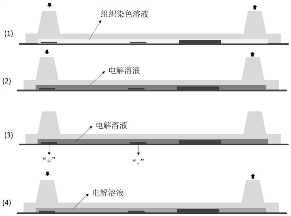
The invention provides a chip device for in-situ tissue dyeing and decoloring. The chip device comprises a three-layer structure which is stacked together and sealed with one another, and the three-layer structure comprises a top cover, a substrate and a bottom plate from top to bottom; the top cover is provided with a liquid inlet and a liquid outlet, one side of the bottom surface of the top cover is concavely provided with a groove, and two ends of the groove are communicated with the liquid inlet and the liquid outlet; a groove corresponding to the bottom surface of the top cover is formed in the substrate and is used as a microfluidic channel; the microfluidic channel is divided into a liquid inlet area, a central area, a tissue fixing area and a liquid outlet area from left to right; a positive electrode and a negative electrode are arranged on the bottom plate, and each electrode is composed of an electrode body and an electrode pin; each electrode body is exposed in the microfluidic channel, and the electrode body of the positive electrode corresponds to the liquid inlet; and the electrode body of the negative electrode is located in the central area. According to internal operation of the chip device, impurity pollution is avoided, the accuracy of analysis is improved, decoloration is conducted through an electrochemical means, consumed time is short, and cell damage is avoided; and the chip device has the characteristic of visualization and is convenient for imaging analysis.

Description

technical field [0001] The invention relates to a chip device for in situ tissue staining and decolorization and a use method thereof, belonging to the technical field of medical inspection and analysis. Background technique [0002] Multiplex Immunohistochemistry (mIHC) uses the principles of immunology and cytochemistry to simultaneously or successively use different colors of fluoresceins or enzymatic products on the same slice to trace tissue or cells in situ. A multi-label staining technique for different antigenic macromolecules. [0003] Multicolor immunohistochemistry technology has emerged along with precise tumor immunotherapy, which can directly evaluate the phenotype and their spatial relationship of various cells in the tumor and tumor microenvironment, reflecting the immune status of the tumor microenvironment. This technology can save time and utilize rare tissue samples to a greater extent, and can help researchers comprehensively, deeply and accurately unde...

Claims


Application Information

Patent Timeline
Patent Type & Authority Applications(China)
IPC IPC(8): G01N1/31B01L3/00
CPCG01N1/31B01L3/5027
Inventor 吴丽苏雪贺月杨马骏秦玉岭
Owner NANTONG UNIVERSITY
Learn More