Pincer-type lower tibiofibular syndesmosis stability detector

A stable and detector technology, applied in the field of medical devices, can solve the problems of increasing patient bleeding and surgical risk, errors in distance measurement and collection, and no objective quantitative detection, etc., to reduce surgical bleeding and surgical risk, reduce radiation, Avoid the effects of fluoroscopy and fibula drilling

Inactive Publication Date: 2020-07-14
青岛山大齐鲁医院(山东大学齐鲁医院(青岛))
View PDF16 Cites 2 Cited by
  • Summary
  • Abstract
  • Description
  • Claims
  • Application Information

AI Technical Summary

Problems solved by technology

[0006] Although the previous detection method of wire tapping test angle has many advantages, there are still the following deficiencies: (1) Although direct radiation exposure can be avoided, corresponding fluoroscopic imaging equipment and microscopic fluoroscopic radiation are still required; It needs to be judged indirectly through the fluoroscopy image; (3) Since the magnification of the fluoroscopy image is affected by the distance between the probe and the object to be seen, there is a certain error in the distance measurement and acquisition; (4) It is necessary to drill a hole on the fibula at the detection site, which is invasive Detection method; (5) X-ray operation takes time, which prolongs the operation time and increases the bleeding and operation risk of the patient; (6) The position and direction of the fibula drilling selection are offset, which may cause the wire tap to resist the center of the lateral edge of the tibia when it enters. As a result, it slips when the stress is applied and loses its diagnostic significance; (7) patients with severe osteoporosis cannot bear the separation stress of the tip of the tap due to their weak bones, so this method cannot be applied; (8) the stress of the test is provided by the progressive force of the tap thread , the tester subjectively estimates the magnitude of the applied stress with the help of hand feeling and experience, without objective quantitative detection

Method used


Image

  • Pincer-type lower tibiofibular syndesmosis stability detector
  • Pincer-type lower tibiofibular syndesmosis stability detector
  • Pincer-type lower tibiofibular syndesmosis stability detector

Examples


Embodiment Construction

[0030] In order to make it easier for those skilled in the art to understand the present invention, the specific implementation manners of the present invention will be described below with reference to the accompanying drawings.

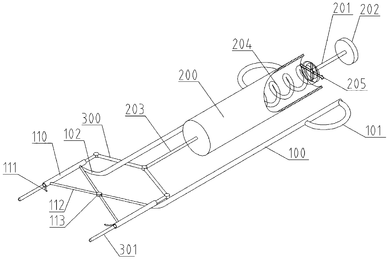
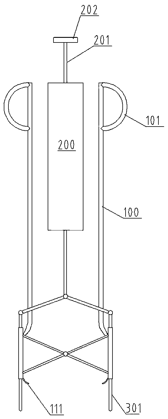
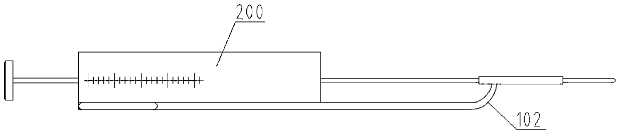
[0031] Such as Figure 1~3 As shown, the present invention provides a clamp-type inferior tibiofibular syndesmosis stability detector, which is characterized in that it includes a fibula holding assembly, a pushing and mechanical measuring assembly, and a separation distance measuring assembly, and the fibula holding assembly includes left and right handles 100 , left and right connecting parts 102 , left and right clamping tubes 110 . The end of the handle 100 is provided with a finger ring 101 to facilitate the opening of the handle 100, the plane where the left and right handles 100 are located is set below the plane where the left and right clamping tubes 110 are located, and the plane where the left and right handles 100 are located The plane ...


PUM


Abstract

The invention relates to the technical field of medical equipment, in particular to a pincer-type lower tibiofibular syndesmosis stability detector. The detector comprises a fibula holding assembly, apushing and mechanics measuring assembly, and a separation distance measuring assembly, wherein the fibula holding assembly comprises handles, connecting parts and clamping pipes, the handles are connected with the clamping pipes through the connecting parts, the clamping pipes are arranged in a hollow structure, and the clamping pipes are movably connected by a connecting mechanism to form a pincer-shaped structure, the pushing and mechanics measuring assembly comprises a force measuring cylinder, a piston rod and a spring, a pressure scale is arranged on the side wall of the force measuringcylinder, the separation distance measuring assembly comprises measuring rods, the measuring rods pass through the clamping pipes, and a ruler is arranged on each measuring rod. The detector enablesthe detection and diagnosis of the lower tibiofibular syndesmosis stability to be based on evidence, and enables a received-force size and separation distance of the lower tibiofibular syndesmosis tobe observed directly and concretely, so that the objectivity and accuracy of the lower tibiofibular syndesmosis stability detection are significantly improved.

Description

technical field [0001] The invention relates to the technical field of medical devices, in particular to a clamp-type inferior tibiofibular syndesmosis stability detector. Background technique [0002] Ankle fracture is a relatively common clinical fracture, accounting for about 3.9% of systemic fractures, ranking first among intra-articular fractures. The main mechanism of injury is rotational violence. Severe ankle joint injuries are often accompanied by separation of the tibiofibular syndesmosis, which accounts for about 1 / 7 of ankle joint injuries. The inferior tibiofibular syndesmosis plays an important role in maintaining the stability of the ankle joint. If it is not diagnosed in time and treated improperly, chronic pain, instability and traumatic arthritis of the ankle joint will often be left behind, which will seriously affect the functional activities of the ankle joint. [0003] The inferior tibiofibular syndesmosis is a ring-shaped structure composed of the dis...

Claims


Application Information

Patent Timeline
Patent Type & Authority Applications(China)
IPC IPC(8): A61B90/00
CPCA61B90/06A61B2090/064
Inventor 赛佳明
Owner 青岛山大齐鲁医院(山东大学齐鲁医院(青岛))
Why Eureka
Try Eureka