
Image processing method for obtaining corneal vertexes

A corneal vertex and image processing technology, applied in the field of image processing, can solve problems such as detection errors, difficulty in fixing the fixed point of the human eye, and large deviations

Active Publication Date: 2020-07-28
WENZHOU MEDICAL UNIV
View PDF4 Cites 5 Cited by
  • Summary
  • Abstract
  • Description
  • Claims
  • Application Information

AI Technical Summary

Problems solved by technology

The measurement accuracy of this method is limited by the diameter of the ring. The larger the diameter of the ring, the lower the accuracy, and it cannot really project to the corneal apex. When the corneal apex and its surroundings are of irregular shape, the center of the ring and The deviation of the true corneal apex will be larger
The Scheimpflug imaging technology uses slit light to irradiate the eye vertically, and obtains the anterior segment section image from the side, so as to obtain the corneal height information to determine the corneal apex. resulting in a certain detection error

Method used


Image

  • Image processing method for obtaining corneal vertexes
  • Image processing method for obtaining corneal vertexes

Examples


Embodiment Construction

[0031] The present invention will be further described below in conjunction with specific embodiments. The embodiments of the present invention are intended to enable those skilled in the art to better understand the present invention, but do not limit the present invention in any way.

[0032] (1): Control the camera to continuously collect n frames of image sequences at high speed, n≥3, subtract the image of the nth frame and the first frame, and calculate whether the image has displacement, because the corneal apex is calculated by this method, and the image needs to be as clear as possible. There is no smearing phenomenon caused by the arrival of the eyes, so the continuous acquisition method ensures that the image has no displacement. For example:

[0033] ∑|f1(x,y)-fn(x,y)|

[0034] or ∑(f1(x,y)-fn(x,y)) 2

[0035] Among them, f1(x, y), fn(x, y) respectively represent the pixel values ​​at the coordinates (x, y) in the first frame and the nth frame of the i...


PUM


Abstract

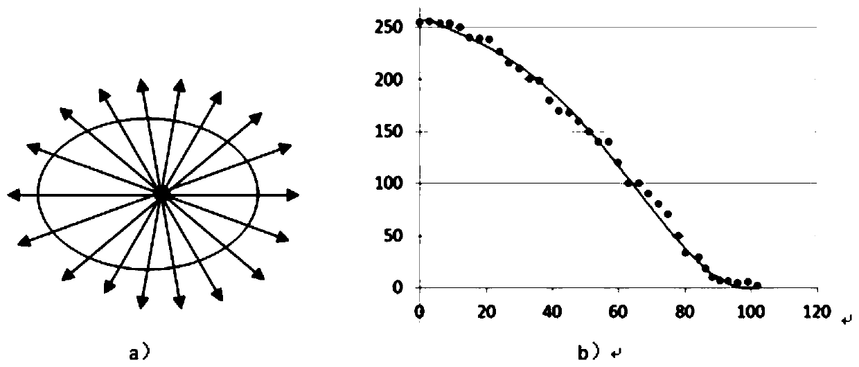
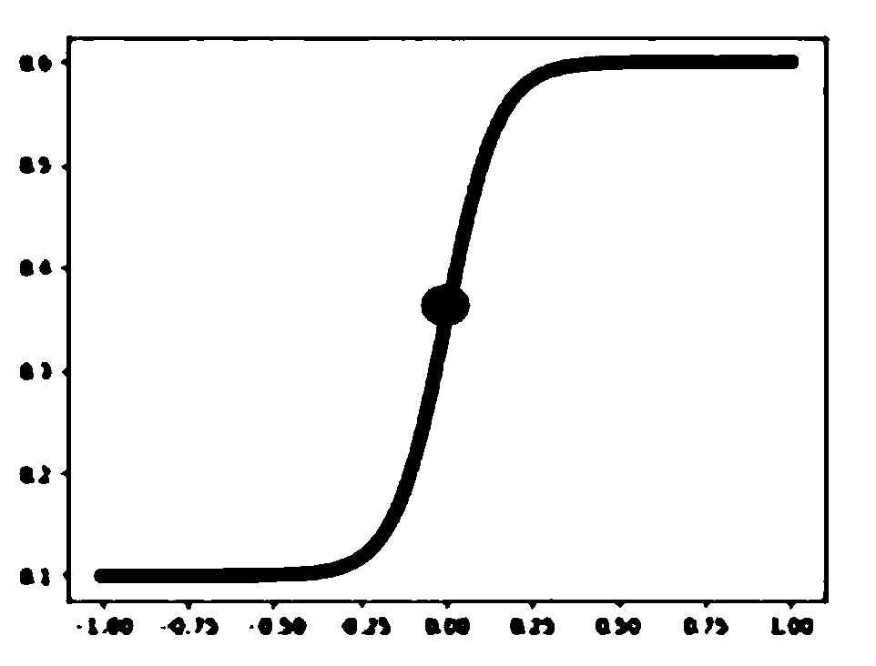
The invention discloses an image processing method for obtaining corneal vertexes. The method comprises the following steps of continuously acquiring N frames of images at a high speed to synthesize aframe of image with minimum noise; acquiring a potential corneal reflection area through image binaryzation and blob analysis so that a to-be-processed area is reduced; carrying out gray stretchingon the potential light spot area; carrying out double-threshold binarization and blob analysis; obtaining a gray gravity center and a light spot boundary coordinate of a cornea vertex area; taking a gray vertex as a center; pulling the ray outwards every a certain angle to obtain a gray sequence on the ray; carrying out gaussian fitting based on a nonlinear least square method on gray scale data of the strip ray group to acquiresub-pixel coordinate values of corneal vertexes; finally, according to the jump of the gray value on the ray at the boundary; solving a sub-pixel-level coordinate boundary; corneal vertex and light spot boundary information obtained through the method can be combined with pupil center position and other related information obtained through other devices to obtain Kappa angle Alpha angle and other related information of eyes, and reliable basic data are provided for subsequent further eye refraction parameter obtaining.

Description

technical field [0001] The invention relates to the technical field of image processing, in particular to an image processing method for obtaining corneal vertices. Background technique [0002] The Kappa angle is the angle between the visual axis and the pupillary axis. Normal people generally have a positive Kappa angle of about 5°. Since the visual axis is a theoretical line, it cannot be directly measured clinically. Therefore, when various instruments measure the Kappa angle, they use the distance between the point of fixation on the cornea and the center of the pupil on the corneal plane to express it, that is, the angular distance between the line of sight and the pupil axis on the corneal plane. There are many measurement methods for Kappa angle. The earlier measurement methods mainly include corneal light reflection method, perimetry, synopic machine, etc., but the measurement accuracy of these methods is very low. The Kappa angle measurement methods currently used...

Claims


Application Information

Patent Timeline
Patent Type & Authority Applications(China)
IPC IPC(8): G06T7/136G06T5/00G06T5/40
CPCG06T7/136G06T5/40G06T2207/30041G06T5/70Y02P90/30
Inventor 赵云娥黄锦海于航
Owner WENZHOU MEDICAL UNIV
Learn More