Deep learning segmentation method based on pseudo-color CT image

A CT image and deep learning technology, applied in the field of medical image processing, can solve problems such as difficult parallel segmentation, improve accuracy and efficiency, and benefit clinical diagnosis and treatment

Pending Publication Date: 2020-08-04
镇江慧影科技发展有限公司
View PDF4 Cites 1 Cited by
  • Summary
  • Abstract
  • Description
  • Claims
  • Application Information

AI Technical Summary

Problems solved by technology

However, for CT images, as well as for different tissues with large differences in observation window width and level, effective observation cannot be performed under the same window width and level, and it is difficult to achieve accurate parallel segmentation

Method used


Image

  • Deep learning segmentation method based on pseudo-color CT image
  • Deep learning segmentation method based on pseudo-color CT image
  • Deep learning segmentation method based on pseudo-color CT image

Examples


Embodiment Construction

[0018] Below in conjunction with accompanying drawing, technical scheme of the present invention is described in further detail:

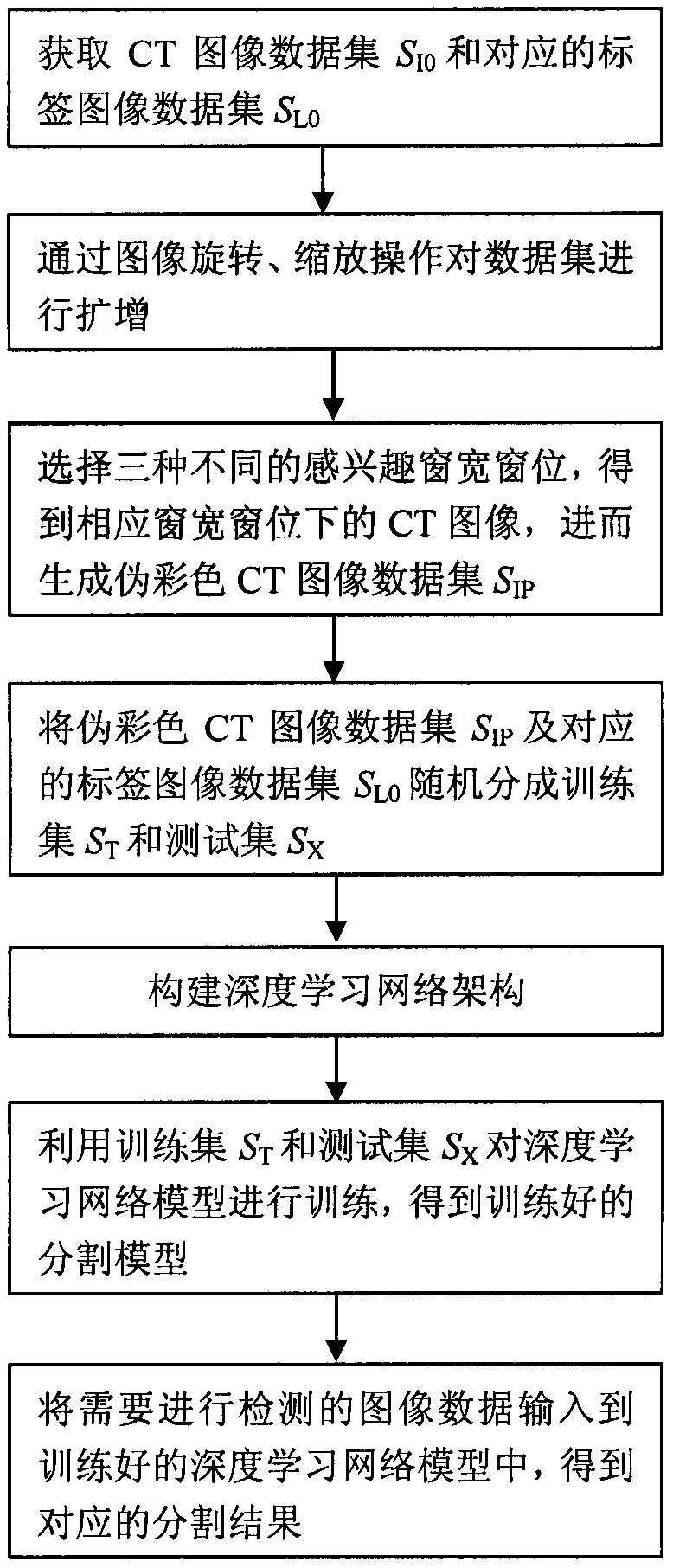
[0019] like figure 1 As shown, the present invention discloses a deep learning segmentation method based on pseudo-color CT images. The following is an example of chest CT image segmentation. The specific steps of segmentation are as follows:

[0020] Step 1: Obtain chest CT image dataset S I0 and the corresponding labeled image dataset S L0 ;

[0021] Step 2: Amplify the data set through image rotation and scaling operations;

[0022] Step 3: Perform windowing processing on the CT image to obtain images under the lung window, heart window, and mediastinal window respectively, and generate a pseudo-color CT image dataset S IP ;

[0023] Step 4: The pseudo-color CT image dataset S IP and the corresponding label image dataset S L0 Randomly divided into training set S T and the test set S X ;

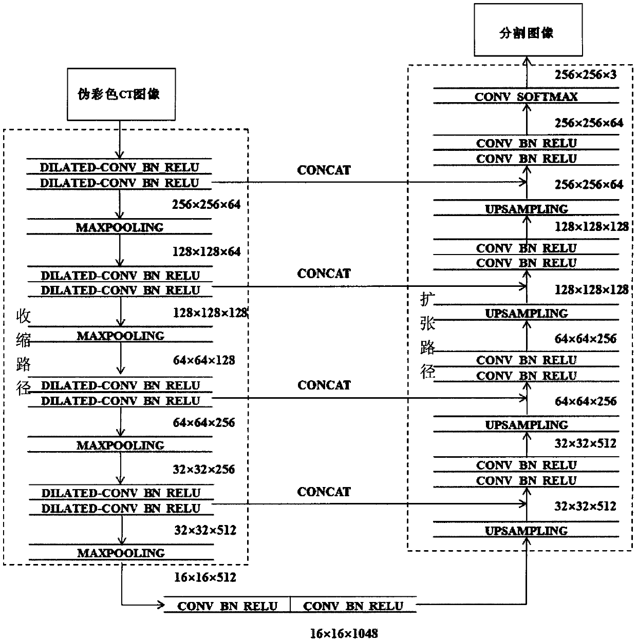
[0024] Step 5: Build a deep learning network ar...


PUM


Abstract

The invention discloses a deep learning segmentation method based on a pseudo-color CT image. The method comprises the following steps: step 1, acquiring a CT image data set SI0 and a corresponding label image data set SL0; 2, amplifying the data set through image rotation and zooming operation; 3, selecting three different interested window width window levels to obtain CT images under the corresponding window width window levels, and further generating a pseudo-color CT image data set SIP; 4, randomly dividing the pseudo-color CT image data set SIP and the corresponding label image data setSL0 into a training set ST and a test set SX; 5, constructing a deep learning network architecture; 6, training the deep learning network model by using the training set ST and the test set SX to obtain a trained segmentation model; and step 7, inputting image data needing to be detected into the trained deep learning network model to obtain a corresponding segmentation result. The CT image segmentation precision and efficiency can be effectively improved, and clinical diagnosis and treatment are facilitated.

Description

technical field [0001] The invention relates to the field of medical image processing, in particular to a deep learning segmentation method based on pseudo-color CT images. Background technique [0002] X-ray CT (Computed Tomography) has the advantages of fast imaging speed and high spatial resolution, and has been widely used in the fields of medical diagnosis and treatment. CT image segmentation is of great significance in 3D reconstruction, computer-aided diagnosis and treatment, radiation therapy and other fields. At present, the segmentation of the tissue of interest mostly depends on the manual outline of the doctor, but the window width and level of observing different tissue images are often quite different, and it is difficult to segment them at the same window width and level at the same time, resulting in image segmentation that not only requires a lot of time Time and energy, and easily affected by subjectivity and doctor experience, there are problems such as i...

Claims


Application Information

Patent Timeline
Patent Type & Authority Applications(China)
IPC IPC(8): G06K9/34G06K9/62G06T11/00
CPCG06T11/008G06V10/267G06F18/214
Inventor 不公告发明人
Owner 镇江慧影科技发展有限公司
Why Eureka
Try Eureka