Method for promoting cartilage repair based on tissue engineering cartilage constructed by co-culture system

A technology for tissue engineering and cartilage repair, applied in the culture process, tissue regeneration, tissue culture, etc., can solve the problems of inducible factor concentration, action time and environment that are difficult to control, prone to hypertrophy or calcification, and poor mechanics of fibrocartilage. Achieve good chondrogenic differentiation potential, good cartilage repair potential, and good biocompatibility

Inactive Publication Date: 2020-08-11
NANJING DRUM TOWER HOSPITAL
View PDF8 Cites 1 Cited by
  • Summary
  • Abstract
  • Description
  • Claims
  • Application Information

AI Technical Summary

Problems solved by technology

However, the directional differentiation regulation mechanism of mesenchymal stem cells is complicated, and the biological behavior of cells is affected by the surrounding microenvironment. It is not enough to simply add one or several induction factors, and the concentration, action time and The environment is difficult to control
In vivo and in vitro results show that the induced mesenchymal stem cells are prone to hypertrophy or calcification, and...

Method used


Image

  • Method for promoting cartilage repair based on tissue engineering cartilage constructed by co-culture system
  • Method for promoting cartilage repair based on tissue engineering cartilage constructed by co-culture system
  • Method for promoting cartilage repair based on tissue engineering cartilage constructed by co-culture system

Examples


Embodiment 1

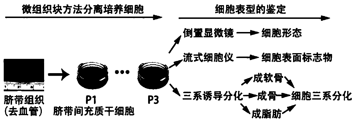
[0040] 1. Separation and acquisition of seed cells

[0041] Isolation and culture of human umbilical cord mesenchymal stem cells: obtain fresh full-term healthy human umbilical cord tissue, clean blood stains, arteriovenous vessels, fascia and adipose tissue under aseptic conditions, obtain Wharton's glue tissue block, cut with sterile scissors Crush the size of 1*1*1mm micro-tissue pieces, add 5ml serum-containing medium (10% FBS, DMEM / F12, 1% glutamine, 1% Vitamin C, 1% penicillin and 1% streptomyces) to the petri dish Vegetarian), place a thermostat (5% CO 2 , 37℃) Isolate umbilical cord mesenchymal stem cells, use micro-tissue block culture method to isolate and culture human umbilical cord mesenchymal stem cells, and pass them to P3 generation cells for the construction of a co-culture system. The cells are fibrous cells under an inverted microscope 状morphology. See the procedures for the isolation, culture and identification of human umbilical cord tissue for umbilical cord...

Embodiment 2

[0048] 1. Isolation, culture and identification of human umbilical cord mesenchymal stem cells (hWJMSCs)

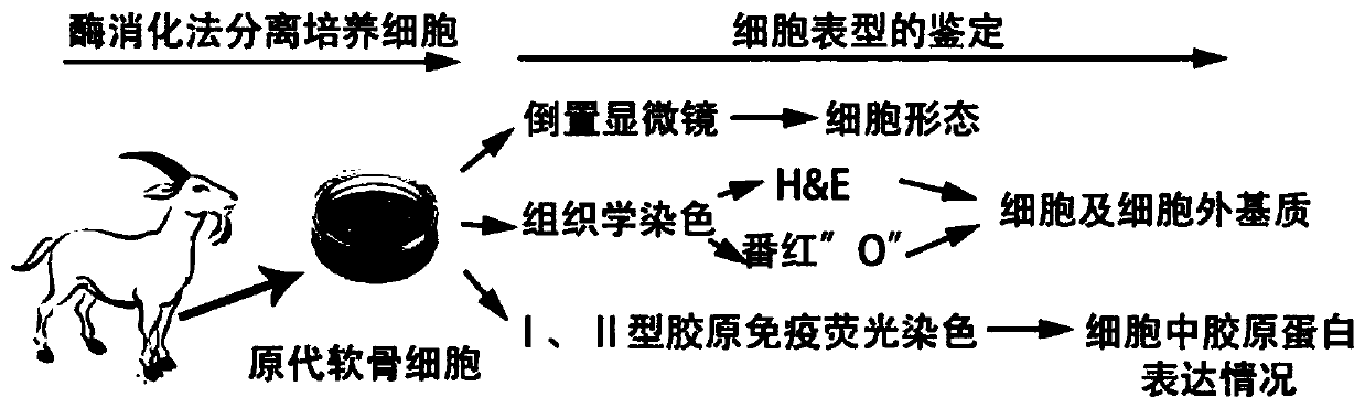
[0049] Fresh full-term healthy human umbilical cords came from the obstetrics department of our hospital. After the patient’s informed consent, the blood was removed under aseptic conditions and then placed in a DMEM / F12 medium culture dish to remove arteriovenous vessels. Wash again several times with culture medium and cut to 1mm 3 Large and small tissue pieces are inoculated into a petri dish with a diameter of 15 cm at an appropriate density, added with 5.0 ml DMEM / F12 (containing 10% fetal bovine serum) medium, and placed in an incubator at 37° C. and 5% CO2 saturated humidity. Add 10.0ml of the same medium the next day, continue to culture for 3 to 5 days to change the medium, and remove the tissue blocks after 10 to 14 days. When the cells adhere to 80% and fuse, the cells are digested with 0.25% trypsin-EDTA to collect the cells, and passaged to the third generation ...


PUM


Abstract

The invention relates to a method for promoting cartilage repair based on tissue engineering cartilage constructed by a co-culture system. The method comprises the following steps: obtaining a small amount of cartilage tissue from a non-weight-bearing area of a knee joint, and separating and culturing primary chondrocytes; separating and culturing umbilical cord mesenchymal stem cells from Wharton's jelly in fresh full-term healthy umbilical cord tissue; performing wet grinding for decellularization by adoption of healthy adult pig knee cartilage tissue, and performing freeze-drying in a vacuum negative pressure environment to obtain an acellular cartilage extracellular matrix oriented scaffold; mixing the primary chondrocytes with the umbilical cord mesenchymal stem cells, planting the cells in a dry scaffold carrier by a cell pendant drop method, performing incubation to construct co-culture tissue, and transplanting the co-culture tissue to full-thickness cartilage defect in the weight-bearing area of the knee joint for articular cartilage repair.

Description

Technical field [0001] The invention relates to the technical field of medical cartilage tissue engineering, in particular to a method for promoting cartilage repair by tissue engineering cartilage constructed based on a co-culture system. Background technique [0002] Due to the lack of blood vessels and lymphatic vessels, cartilage tissue can hardly heal itself once it is damaged. As time goes by, it will be secondary to osteoarthritis. At present, there is no effective clinical strategy to repair or reverse the damaged cartilage. Tissue engineering combines seed cells, biomaterials and cytokines to construct tissue engineered cartilage, bringing new hope for cartilage repair and regeneration. However, the source of chondrocytes is limited. For large area cartilage defects, it is almost impossible to provide sufficient cartilage tissue for the isolation and extraction of chondrocytes. In vitro expansion is extremely prone to dedifferentiation and lose phenotype. Mesenchymal ste...

Claims


Application Information

Patent Timeline
IPC IPC(8): A61L27/36A61L27/38C12N5/077C12N5/0775
CPCA61L27/3612A61L27/3654A61L27/3817A61L27/3834A61L27/3852A61L27/3886A61L2430/06C12N5/0655C12N5/0668C12N2500/32C12N2500/38C12N2509/00
Inventor 张雨蒋青陈东阳秦江辉
Owner NANJING DRUM TOWER HOSPITAL
Why Eureka
Try Eureka