Protein electrophoresis staining solution, kit and staining method
A dyeing solution and protein technology, which is applied in biological testing, preparation of test samples, material analysis by electromagnetic means, etc., can solve the problems of long dyeing and decolorization, and achieve shortened dyeing time, low background dyeing, and dyeing speed. quick effect
- Summary
- Abstract
- Description
- Claims
- Application Information
AI Technical Summary
Problems solved by technology
Method used
Image
Examples
Embodiment 1
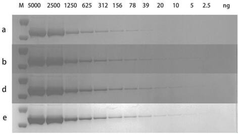
[0048] 1. Preparation of Staining Solution
[0049] Weigh 250 mg Coomassie Brilliant Blue G-250 (CBB G-250), 300 mg Coomassie Brilliant Blue R-250 (CBB R-250), 100 ml glycerin, 1 ml essence, 20 g trehalose, 1 g Tween 20 , add deionized water to make up to 985ml, stir for 2-4 hours, after the Coomassie Brilliant Blue is fully dissolved, add 15ml of concentrated hydrochloric acid with a concentration of 38%, after mixing evenly, the final acidity of the solution is 14. Finally, store it in opaque bottles.
[0050] 2. SDS-PAGE electrophoresis band staining and decolorization
[0051] 1) After protein gel electrophoresis, wash the gel with 100ml of deionized water on a shaker in a plastic container for 1 min, discard the washed water, and repeat twice.
[0052] 2) Add 100ml of deionized water, heat it in a microwave oven to near boiling, but avoid boiling, then shake and wash on a shaker for 5 minutes. For gels with a thickness of 1.5mm, it is necessary to prolong the shaking w...
Embodiment 2
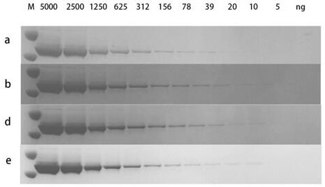
[0060] 1. Preparation of Staining Solution
[0061] Weigh 250mg of Coomassie Brilliant Blue G-250 (CBB G-250), 100ml of glycerin, 1ml of essence, 20g of trehalose, 1g of Tween 20, add deionized water to 985ml, stir for 2 to 4 hours, and wait for examination After the Mas Brilliant Blue is fully dissolved, add 15ml of concentrated hydrochloric acid with a concentration of 38%, and after mixing evenly, the final acidity of the solution is 14. Finally, store it in opaque bottles.
[0062] 2. SDS-PAGE electrophoresis band staining and decolorization
[0063] 1) After protein gel electrophoresis, wash the gel with 100ml of deionized water on a shaker in a plastic container for 1 min, discard the washed water, and repeat twice.
[0064] 2) Add 100ml of deionized water, heat it in a microwave oven to near boiling, but avoid boiling, then shake and wash on a shaker for 5 minutes. For gels with a thickness of 1.5mm, it is necessary to prolong the shaking washing time.
[0065] 3) S...
Embodiment 3
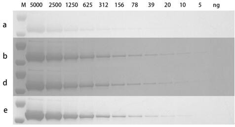
[0072] 1. Preparation of Staining Solution
[0073] Weigh 300 mg Coomassie Brilliant Blue R-250 (CBB R-250), 100 ml glycerin, 1 ml essence, 20 g trehalose, 1 g Tween 20, add deionized water to 985 ml, stir for 2-4 h, After the Coomassie Brilliant Blue is fully dissolved, add 15ml of concentrated hydrochloric acid with a concentration of 38%, and after mixing evenly, the final acidity of the solution is 14. Finally, store it in opaque bottles.
[0074] 2. SDS-PAGE electrophoresis band staining and decolorization
[0075] 1) After protein gel electrophoresis, wash the gel with 100ml of deionized water on a shaker in a plastic container for 1 min, discard the washed water, and repeat twice.
[0076] 2) Add 100 ml of deionized water, heat it in a microwave oven to near boiling, but avoid boiling, then shake and wash on a shaker for 5 minutes. For gels with a thickness of 1.5mm, it is necessary to prolong the shaking washing time.
[0077] 3) Shake and wash the gel with 100ml o...
PUM
| Property | Measurement | Unit |
|---|---|---|
| Bronsted acidity | aaaaa | aaaaa |
| Bronsted acidity | aaaaa | aaaaa |
Abstract
Description
Claims
Application Information
 - R&D
- Intellectual Property
- Life Sciences
- Materials
- Tech Scout
- Unparalleled Data Quality
- Higher Quality Content
- 60% Fewer Hallucinations
Browse by: Latest US Patents, China's latest patents, Technical Efficacy Thesaurus, Application Domain, Technology Topic, Popular Technical Reports.
© 2025 PatSnap. All rights reserved.Legal|Privacy policy|Modern Slavery Act Transparency Statement|Sitemap|About US| Contact US: help@patsnap.com


