Supercharge Your Innovation With Domain-Expert AI Agents!

Multi-modal medical image conversion method based on artificial intelligence

A technology of medical imaging and conversion method, applied in the field of medical imaging, can solve the problems of speckle noise interference, penetration, difficult automatic segmentation of OCT images, etc., and achieve the effect of solving edge blurring

Pending Publication Date: 2020-09-08
中国医学科学院阜外医院深圳医院
View PDF5 Cites 5 Cited by
  • Summary
  • Abstract
  • Description
  • Claims
  • Application Information

AI Technical Summary

Problems solved by technology

However, considering the complex anatomical structures of false lumen, true lumen and thrombus in thoracic aortic dissection, and due to the characteristics of the three-layer tissue structure of the arterial wall, there will be backscatter in OCT imaging, resulting in signal" The attenuation and penetration of the OCT image, and the interference of speckle noise at the same time, eventually form a weak edge phenomenon, which brings difficulties to the automatic segmentation of OCT images.

Method used


Image

  • Multi-modal medical image conversion method based on artificial intelligence
  • Multi-modal medical image conversion method based on artificial intelligence
  • Multi-modal medical image conversion method based on artificial intelligence

Examples


Embodiment 1

[0042] Embodiment 1: A method for converting multimodal medical images based on artificial intelligence

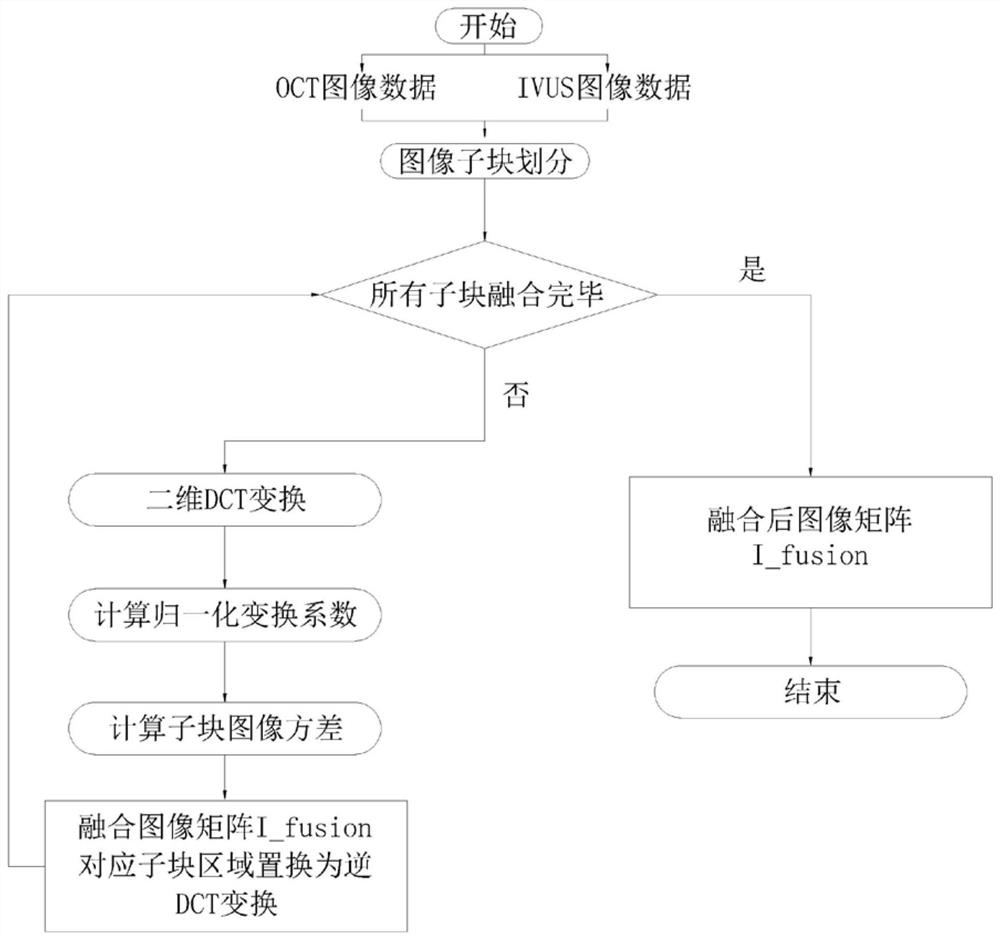
[0043] Simply using the single grayscale information of the OCT image can only segment the outer wall and inner wall of the artery, but it cannot clearly describe the details of the middle part of the artery from the intima to the adventitia, that is, it cannot accurately segment the false lumen and The border of the thrombus. Therefore, this example solves the problem through multimodal information fusion.

[0044] It specifically includes the following steps:

[0045] 1) Obtain OCT image data of aortic dissection

[0046] Ex vivo imaging of vessels in thoracic aortic dissection using optical coherence tomography (OCT),

[0047] OCT imaging technology is currently the highest resolution intravascular imaging technology, which can effectively display the inner wall structure of blood vessels, identify the tearing of vascular intima, plaque tissue components, and poor st...


PUM


Abstract

The invention discloses a multi-modal medical image conversion method based on artificial intelligence, and mainly relates to the field of medical images. The method comprises the following steps: acquiring OCT image data of aortic dissection; acquiring IVUS image data of the aortic dissection; a global threshold segmentation method is adopted for the background of the OCT image data and the background of the IVUS image data, the outer contour of the aorta is extracted, and morphological hole filling and area threshold methods are used for cleaning noise information; and performing image information fusion on the OCT image data and the IVUS image data through a discrete cosine transform frequency domain information fusion method to obtain a fused image. The beneficial effects of the invention are that the method combines thoracic aortic dissection images of various modes, enables the structural features in an ultrasonic image to be fused into an OCT image, and effectively improves thefeatures of a true cavity, a false cavity and a thrombus in the fused multi-mode image.

Description

technical field [0001] The invention relates to the field of medical imaging, in particular to an artificial intelligence-based multimodal medical image conversion method. Background technique [0002] In recent years, with the rapid development of various medical imaging technologies (CT.MRI.Utrasound.PET, OCT, Microscop, etc.), and have been widely used in the early detection, diagnosis and treatment of diseases, resulting in clinical, imaging Experts need to face a large amount of medical imaging data of patients every day. Simply relying on manual reading and judgment of these imaging data is not only time-consuming and laborious, but also subjective. Therefore, computer-aided diagnosis technology has become a powerful measure to solve this problem. In particular, the introduction of a machine learning method "deep learning" that has emerged in recent years has gradually accelerated the pace of modern computer medical image computing and analysis becoming intelligent an...

Claims


Application Information

Patent Timeline
IPC IPC(8): G06T7/00G06T7/136G06T7/194G06T5/50
CPCG06T7/0012G06T7/136G06T7/194G06T5/50G06T2207/10081G06T2207/10101G06T2207/10104G06T2207/10132G06T2207/20052G06T2207/20192G06T2207/20221G06T2207/30101
Inventor 孙凯袁旭春高立蔡震宇李亿华李涯
Owner 中国医学科学院阜外医院深圳医院
Features
  • R&D
  • Intellectual Property
  • Life Sciences
  • Materials
  • Tech Scout
  • Unparalleled Data Quality
  • Higher Quality Content
  • 60% Fewer Hallucinations
Learn More