
Ultra-fast ultrasonic Doppler spinal cord micro-blood-flow imaging system

A Doppler spinal cord and imaging system technology, which is applied in blood flow measurement devices, ultrasonic/sonic/infrasonic diagnosis, and the structure of ultrasonic/sonic/infrasonic diagnostic equipment, can solve the problem of long imaging time, high cost and human health. Harmful and other problems, to achieve the effect of improving resolution and signal-to-noise ratio

Active Publication Date: 2020-10-16
FUDAN UNIV
View PDF5 Cites 3 Cited by
  • Summary
  • Abstract
  • Description
  • Claims
  • Application Information

AI Technical Summary

Problems solved by technology

These methods still have the following shortcomings: there is electromagnetic radiation harmful to human health, the cost is high, and the imaging time is long

Method used


Image

  • Ultra-fast ultrasonic Doppler spinal cord micro-blood-flow imaging system
  • Ultra-fast ultrasonic Doppler spinal cord micro-blood-flow imaging system
  • Ultra-fast ultrasonic Doppler spinal cord micro-blood-flow imaging system

Examples


Embodiment Construction

[0051] In order to make the object, technical solution and advantages of the present invention clearer, the present invention will be described in detail below in conjunction with the embodiments and accompanying drawings. The specific embodiments described here are only used to explain the present invention, not to limit the present invention.

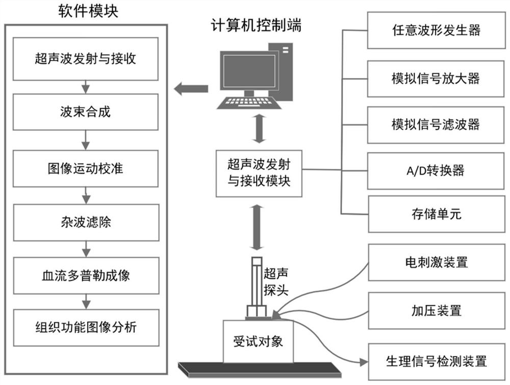
[0052] figure 1 It is the spinal cord micro-flow ultrasonic imaging system proposed by the present invention, and the system block diagram is also applicable to this embodiment. This embodiment uses the spinal cord micro-flow imaging system proposed by the present invention to image the rat spinal cord, but is not limited to the imaging of the rat spinal cord.

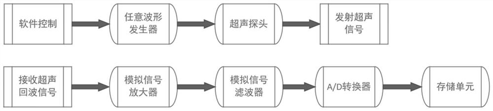
[0053] 1. Design the ultrasonic transmission and reception sequence

[0054] In the present invention, ultrafast ultrasonic plane wave imaging technology is used to perform high frame rate and high quality imaging of spinal cord micro blood flow. Therefore, first of all, i...


PUM


Abstract

The invention belongs to the technical field of ultrasonic imaging of biomedicine and particularly relates to an ultra-fast ultrasonic Doppler spinal cord micro-blood-flow imaging system. The system disclosed by the invention comprises two parts, i.e., hardware and software, wherein the hardware part comprises an ultrasonic wave transmitting and receiving module and an experimental apparatus module; and the software part comprises modules of ultra-fast ultrasonic imaging, wave beam synthesizing, movement calibrating, clutter filtering, Doppler imaging, differencing and relevance analyzing. Firstly, an ultrasonic plane-wave transmitting and receiving control module is compiled based on an ultra-fast ultrasonic imaging technique and a multiangular plane-wave integrating imaging theory, and hardware units are controlled to transmit and receive ultrasonic waves by computer software; received echo data are processed, so as to finally obtain a Doppler blood flow image; and parameters such asvelocity and direction of blood flow are analyzed. The system further provides a mode for imaging micro-blood-flow of a spinal cord under the condition that the spinal cord is pressurized, injured and stimulated, and functional analysis and physiological and pathological analysis on the spinal cord can be carried out.

Description

technical field [0001] The invention belongs to the technical field of biomedical ultrasonic imaging, in particular to a spinal cord micro-blood flow imaging system. Background technique [0002] Spinal cord injury is often accompanied by functional nerve injury, and the complete loss of blood flow in the trauma center and the reduction of peripheral blood perfusion can be used to indicate the lesion of spinal cord function injury. [0003] At present, the standard clinical diagnostic methods for spinal injuries include X-ray plain film, computed tomography (CT) and magnetic resonance imaging (MRI). These methods still have the following disadvantages: there is electromagnetic radiation harmful to human health, the cost is high, and the imaging time is long. Compared with these existing methods, ultrasound imaging has the advantages of fast imaging speed, portable equipment, low cost and no ionizing radiation. Ultrafast ultrasonic Doppler blood flow imaging method based on...

Claims


Application Information

Patent Timeline
IPC IPC(8): A61B8/06A61B8/00
CPCA61B8/06A61B8/488A61B8/5269A61B8/44
Inventor 许凯亮臧佳琦闫少渊他得安王威琪
Owner FUDAN UNIV
Learn More