Endoscope imaging method and endoscope imaging system

An imaging method and imaging system technology, applied in endoscopy, medical science, diagnosis, etc., can solve problems such as inconsistency in the clarity of visible light images and fluorescent monochromatic images, and differences in focal lengths.

Pending Publication Date: 2020-10-23
SHENZHEN PROXINSE MEDICAL LTD
View PDF0 Cites 12 Cited by
  • Summary
  • Abstract
  • Description
  • Claims
  • Application Information

AI Technical Summary

Problems solved by technology

Whether it is time-sharing or light-splitting, in the prior art, visible light, return excitation light, and excitation light all share a set of front-end optical links. Due to the differen

Method used


Image

  • Endoscope imaging method and endoscope imaging system
  • Endoscope imaging method and endoscope imaging system
  • Endoscope imaging method and endoscope imaging system

Examples


Embodiment 1

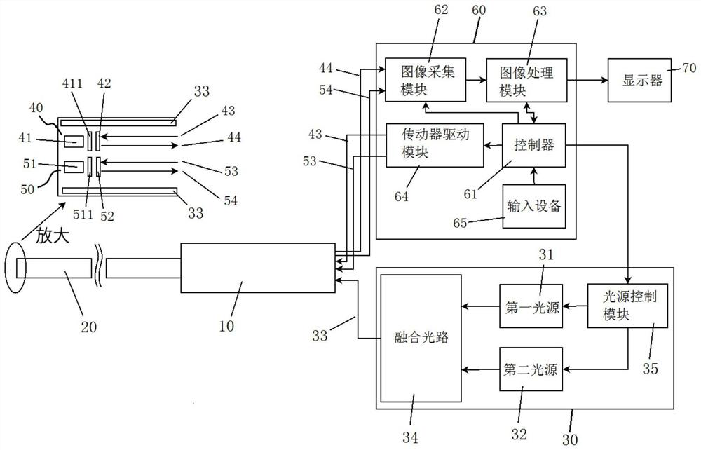
[0042] This embodiment provides an endoscope imaging system. The endoscope imaging system is a binocular endoscope and is mainly used for detecting human cancer.

[0043] Such as figure 1 As shown, the endoscopic imaging system mainly includes a handle 10, a mirror tube 20, a light source assembly 30, a first front end assembly 40, a second front end assembly 50, and a control device 60, and other parts of the endoscopic imaging system are not shown in this presentation. Involved in the application, it will not be described in detail.

[0044] The mirror tube 20 is a hard mirror tube or a soft mirror tube. The rear end of the mirror tube 20 is connected to the front end of the handle 10. The handle 10 is provided with a threading channel communicating with the mirror tube 20. The handle 10 is used for doctors to operate the mirror tube 20. The front end extends into the human body.

[0045]The light source assembly 30 includes a first light source 31, a second light source 3...


PUM


Abstract

The invention discloses an endoscope imaging method and an endoscope imaging system. The endoscope imaging method comprises the following steps of illumination, visible light and fluorescence collection, image signal collection and image processing. Visible light reflected by a human subject and excited fluorescence are collected through two sensors located at the front end of an endoscope tube, corresponding image signals are generated, and then the image signals are transmitted to the rear end to be processed to obtain a final endoscope image. Compared with the prior art that visible light and fluorescence are transmitted to the rear end through a light path and then collected and processed, the endoscope of the invention solves the problem that the precision is reduced due to light pathpropagation, the imaging precision is improved, and the light path cost and the light path occupied space are also saved; in addition, the visible light and the fluorescence can be focused and collected respectively due to the design of independent light paths of the visible light and the fluorescence, so that the visible light image and the fluorescence image are consistent in definition; and the endoscope imaging system is binocular stereoscopic vision, and can obtain three-dimensional information to compensate fluorescence brightness of different depth areas.

Description

technical field [0001] The invention relates to the technical field of medical instruments, in particular to an endoscope imaging method and an endoscope imaging system. Background technique [0002] In minimally invasive surgery, the application of excitation light endoscopy (or fluorescence endoscopy) can accurately locate the location of cancer, and then perform more accurate resection of cancerous tissue. Its main principle is: After dispersing or injecting excitation photopharmaceuticals into the target part of the living body, the excitation light from the light source device is irradiated to the subject, and by capturing the fluorescence from the cancer, the existence diagnosis and malignancy of cancer are carried out. Qualitative diagnosis of the degree and so on. [0003] The prior art is mainly realized by single-camera time-sharing imaging and dual-camera spectroscopic imaging. Spectroscopic technology is mainly used at the handle end to separate white light, ex...

Claims


Application Information

Patent Timeline
IPC IPC(8): A61B1/04
CPCA61B1/00163A61B1/00186A61B1/043
Inventor 陆汇海
Owner SHENZHEN PROXINSE MEDICAL LTD
Why Eureka
Try Eureka