Eureka AIR delivers breakthrough ideas for toughest innovation challenges, trusted by R&D personnel around the world.

Medical image processing system and method and computer storage medium

A medical image and processing system technology, applied in the field of medical image processing systems, can solve the problems of long imaging time, high cost of MR image reconstruction, and dependence.

Pending Publication Date: 2020-10-23
SHANGHAI UNITED IMAGING INTELLIGENT MEDICAL TECH CO LTD
View PDF0 Cites 2 Cited by
  • Summary
  • Abstract
  • Description
  • Claims
  • Application Information

AI Technical Summary

Problems solved by technology

However, due to the slow imaging speed and long imaging time of magnetic resonance imaging technology, some patients will involuntarily move during the imaging process, resulting in motion artifacts, which affect the imaging quality.
[0003] In order to obtain high-quality MR images in a short time, in related technologies, equipment manufacturers will equip MR imaging equipment with hardware equipment, that is, a reconstruction workstation, through which the MR images are reconstructed to obtain better quality reconstructed MR images, but This method of reconstruction of MR images is not only slow in reconstruction speed, but also needs to rely on expensive hardware equipment, resulting in high reconstruction costs of MR images

Method used


Image

  • Medical image processing system and method and computer storage medium
  • Medical image processing system and method and computer storage medium
  • Medical image processing system and method and computer storage medium

Examples


Embodiment Construction

[0073] The technical solutions in the embodiments of the present invention will be clearly and completely described below in conjunction with the accompanying drawings in the embodiments of the present invention. Obviously, the described embodiments are only a part of the embodiments of the present invention, rather than all the embodiments. Based on the embodiments of the present invention, all other embodiments obtained by those of ordinary skill in the art without creative work shall fall within the protection scope of the present invention.

[0074] It should be noted that the terms "first" and "second" in the specification and claims of the present invention and the above-mentioned drawings are used to distinguish similar objects, and not necessarily used to describe a specific sequence or sequence. It should be understood that the data used in this way can be interchanged under appropriate circumstances so that the embodiments of the present invention described herein can be...


PUM


Abstract

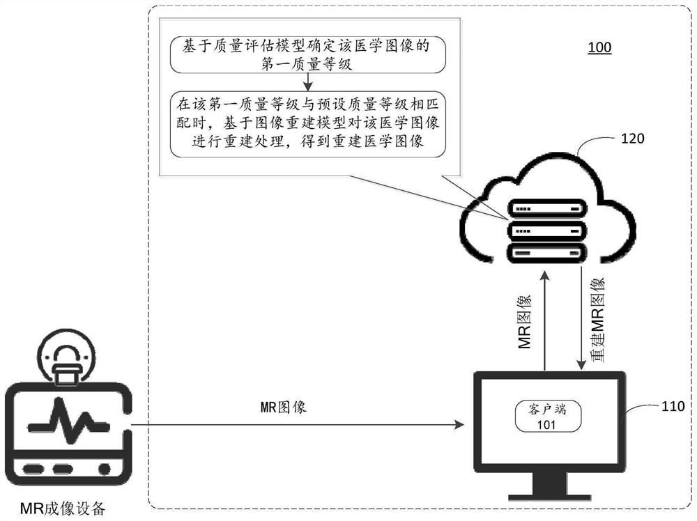
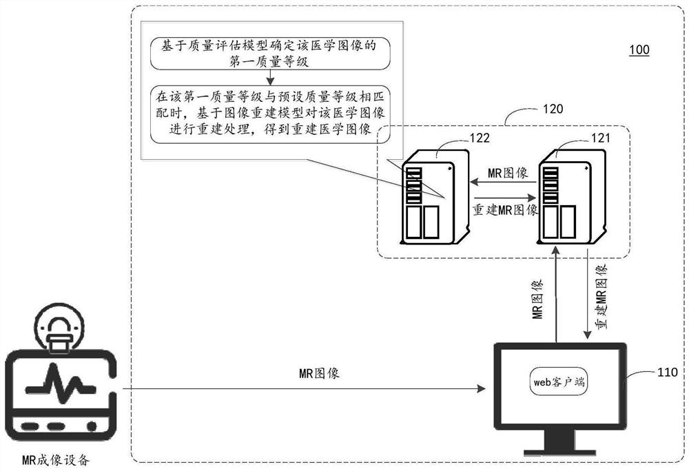
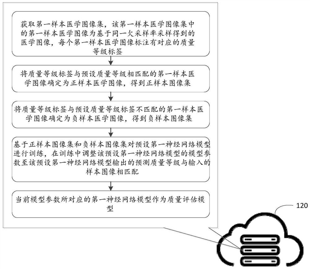
The invention discloses a medical image processing system and method, and a computer storage medium. The system comprises a first terminal which is used for obtaining a medical image from medical imaging equipment, and transmitting the medical image to a cloud server; the cloud server is used for receiving the medical image and determining a first quality grade of the medical image based on a quality evaluation model; when the first quality grade is matched with a preset quality grade, performing reconstruction processing on the medical image based on an image reconstruction model to obtain areconstructed medical image; returning the reconstructed medical image to the first terminal; wherein the quality evaluation model is determined by performing machine learning training based on the first sample medical image and the corresponding quality grade label; wherein the image reconstruction model is determined by performing machine learning training based on a full-sampling medical imageand an under-sampling medical image corresponding to the full-sampling medical image. According to the method, the reconstruction cost of the medical image is reduced, and the reconstruction efficiency and the quality of the reconstructed image are improved.

Description

Technical field [0001] The present invention relates to the technical field of medical image processing, in particular to a medical image processing system, method and computer storage medium. Background technique [0002] Magnetic Resonance (MR) imaging technology is not only ideal in terms of tissue contrast, spatial resolution, etc., but also has good imaging effects on soft tissues and can provide richer image information, so it is widely used. However, the slow imaging speed and long imaging time of magnetic resonance imaging technology cause some patients to move involuntarily during the imaging process, resulting in motion artifacts, which affects the imaging quality. [0003] In order to be able to obtain high-quality MR images in a short time, in related technologies, equipment manufacturers will equip MR imaging equipment with hardware equipment, namely reconstruction workstations, through which MR images can be reconstructed to obtain better quality reconstructed MR imag...

Claims


Application Information

Patent Timeline
Patent Type & Authority Applications(China)
IPC IPC(8): G06T7/00G06T17/00G06N3/04G06N3/08G06K9/62
CPCG06T7/0012G06T17/00G06N3/084G06T2207/10072G06T2207/30168G06N3/045G06F18/22
Inventor 吕旭阳廖术
Owner SHANGHAI UNITED IMAGING INTELLIGENT MEDICAL TECH CO LTD
Why Eureka
Eureka Blog
Learn More