Gland cell image segmentation method and system based on improved U-Net network

An image segmentation, cell technology, applied in image analysis, image enhancement, image data processing and other directions, can solve the problem of limiting the capture of finer details, unable to capture and so on

Pending Publication Date: 2020-12-01
HANGZHOU NORMAL UNIVERSITY
View PDF0 Cites 9 Cited by
  • Summary
  • Abstract
  • Description
  • Claims
  • Application Information

AI Technical Summary

Problems solved by technology

Despite the use of skip connections capable of propagating information from shallow to deep layers, the network fails to capture more detailed information due to the standard encoder-decoder architecture, where the dimensionality of the data is reduced near the bottleneck, the first few Blocks learn low-level features of the data, while later blocks learn high-level features, and eventually, the encoder learns to map the data to lower dimensions (in the spatial sense)
The ever-increasing receptive tiny structures in the network depth require smaller receptive fields, and in the case of standard U-Net, even with skip connections, the smallest receptive field is limited by the first layer
Thus, incomplete architectures inherently limit their ability to capture finer details

Method used


Image

  • Gland cell image segmentation method and system based on improved U-Net network
  • Gland cell image segmentation method and system based on improved U-Net network
  • Gland cell image segmentation method and system based on improved U-Net network

Examples


Embodiment Construction

[0059] The present invention is further analyzed below in conjunction with specific embodiment.

[0060] A glandular cell segmentation method based on the improved U-Net network, including the following:

[0061] Step 1. Data Acquisition

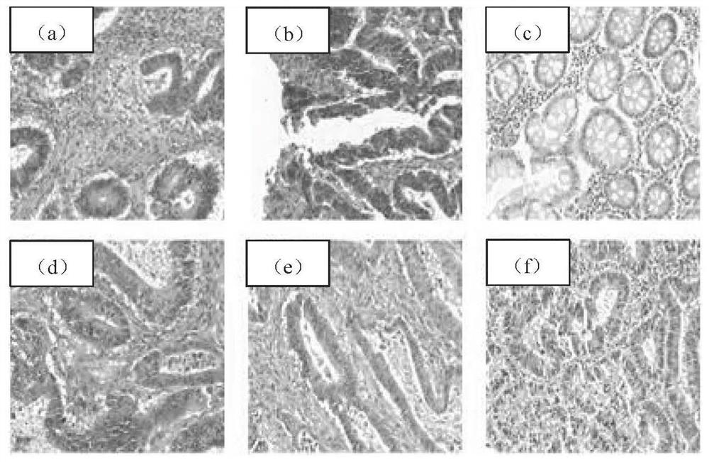
[0062] Obtain figure 1 The original image of glandular cells is generally obtained from related competitions, such as MICCAI2015 Gland Segmentation Challenge dataset (Glas);

[0063] Step 2. Data preprocessing

[0064] 2.1 Since the acceptable image size of the U-Net network is 512x512, the image size is adjusted for the original image of glandular cells.

[0065] 2.2 Since some data have low resolution and uneven staining, images with high resolution and no lack of information (some without staining indicate that they are not cells, so information is missing) were selected as training data.

[0066] 2.3 Use the tensorflow data enhancement library to enhance the data set of the training data set, so that the data of the training data set...


PUM


Abstract

The invention discloses a gland cell image segmentation method and system based on an improved U-Net network. According to the method, the convolution layers are combined to construct the dense connection block, and the U-Net is amplified by a multi-resolution analysis method, so that the problem of cell segmentation with different rules can be well solved. And a self-attention module, namely a position attention module and a channel attention module, is introduced at a decoder end, so that context information can be adaptively aggregated, and the feature representation of cell segmentation isimproved. The cell image is segmented by using the network, so that the segmentation effect is improved. The method is easy to implement, data preprocessing operation is simple, and better robustnessand accuracy are achieved.

Description

technical field [0001] The invention relates to the technical field of network pattern recognition and segmentation, in particular to a non-diagnostic purpose-based glandular cell image segmentation method and system based on a densely connected block U-Net network. Background technique [0002] Histopathological image analysis plays a crucial role for accurate tumor cell estimation and prognosis. The size, shape, and some other morphological manifestations of imaged structures such as nuclei and glands are highly correlated with the presence or severity of disease. Previously, these images were evaluated by pathologists who evaluated biopsy samples, but the manual process was subjective, labor-intensive, and time-consuming. Therefore, computational methods to quantitatively and objectively analyze histopathology images have been developed. Image segmentation, i.e. the extraction of cells, nuclei or glands from histopathological images, is a step preceding all analysis and...

Claims


Application Information

Patent Timeline
Patent Type & Authority Applications(China)
IPC IPC(8): G06T7/11G06T7/00G06N3/04G06N3/08G06K9/62G06F17/16
CPCG06T7/11G06T7/0012G06N3/08G06F17/16G06T2207/30024G06N3/045G06F18/2415G06F18/253G06F18/214
Inventor 赵宝奇孙军梅李秀梅
Owner HANGZHOU NORMAL UNIVERSITY
Why Eureka
Try Eureka