Polypeptide segment diagnosis model and application thereof in prediction of myeloblastoma metastasis risk
A technology of medulloblastoma and peptides, which is applied in the diagnostic model of polypeptide segments and its application in predicting the risk of medulloblastoma metastasis, can solve the problem that differential proteins cannot be found, quantitative analysis stability and sensitivity decline, etc. question
- Summary
- Abstract
- Description
- Claims
- Application Information
AI Technical Summary
Problems solved by technology
Method used
Image
Examples
Embodiment 1
[0018] Embodiment 1, research basis of the present invention
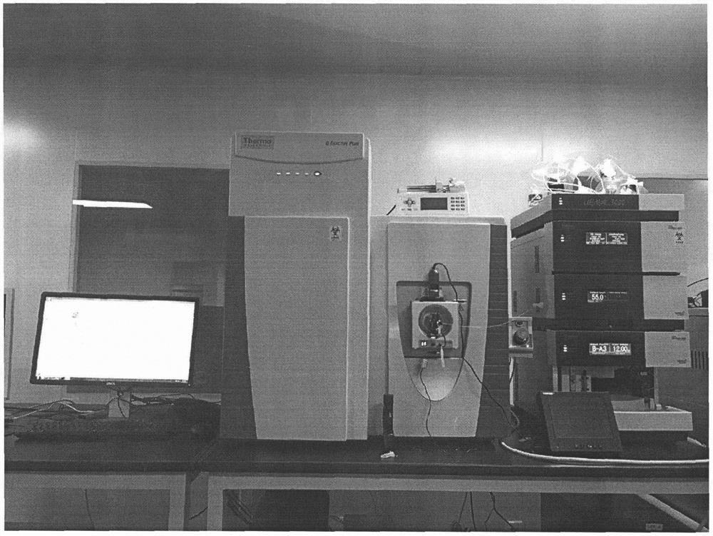
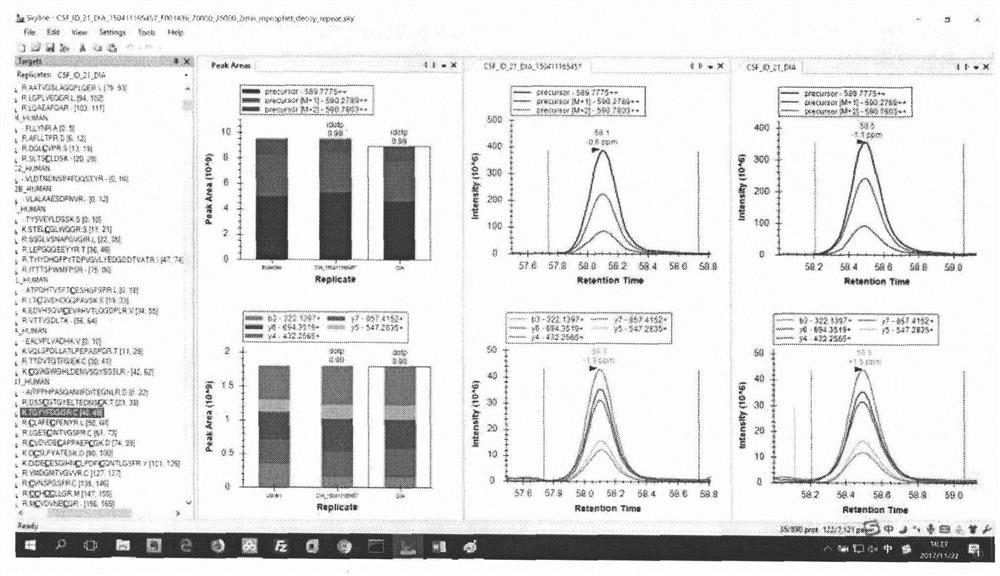
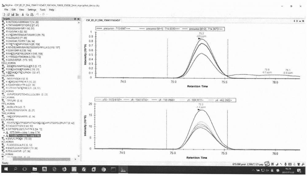
[0019] Analysis of peptide content in cerebrospinal fluid by DIA method
[0020] CSF sample preparation: Cells were centrifuged at 200009 for 10 minutes at 4°C in a refrigerated centrifuge to remove insoluble material and cells. Add 1M DTT to each case of cerebrospinal fluid to a final concentration of 5mM, and reduce at 56°C for 1 hour. After cooling to room temperature, 0.5M IAM was added to a final concentration of 10 mM, and alkylation was carried out at room temperature for 45 minutes. The final concentration of L-cysteine at room temperature was 20mM, and the alkylation reaction was terminated for 20 minutes; the final concentration of 1M TEAB was added to 0.1M, and trypsin (trypsin: sample, 1:50, m / m) was hydrolyzed overnight , add trypsin again for 4 hours the next day; add formic acid to a final concentration of 1% to stop the enzymatic hydrolysis; extract the product with a C18 extraction column, dry ...
Embodiment 2
[0027] Embodiment 2, detection method of the present invention
[0028] 75 peptides that make up the diagnostic model
[0029] 1. CO5_HUMAN_LQGTLPVEAR_2_y7_1
[0030] 2. CO5_HUMAN_LQGTLPVEAR_2_y8_1
[0031] 3. ITIH4_HUMAN_TGLLLLSDPDK_2_precursor_2
[0032] 4. ITIH4_HUMAN_TGLLLLSDPDK_2_precursor[M+1]_2
[0033] 5. PRDX6_HUMAN_LPFPIIDDR_2_precursor[M+1]_2
[0034] 6. 7B2_HUMAN_SVNPYLQGQR_2_precursor_2
[0035] 7. 7B2_HUMAN_SVNPYLQGQR_2_precursor[M+1]_2
[0036] 8. 7B2_HUMAN_SVNPYLQGQR_2_precursor[M+2]_2
[0037] 9. 7B2_HUMAN_SVNPYLQGQR_2_y4_1
[0038] 10. 7B2_HUMAN_SVNPYLQGQR_2_y5_1
[0039] 11. 7B2_HUMAN_SVNPYLQGQR_2_y7_1
[0040] 12. 7B2_HUMAN_SVNPYLQGQR_2_y8_1
[0041] Thirteen, ANGT_HUMAN_VEGLTFQQNSLNWMK_2_y3_1
[0042] Fourteen, ANGT_HUMAN_VEGLTFQQNSLNWMK_2_y4_1
[0043] Fifteen, ANGT_HUMAN_VEGLTFQQNSLNWMK_2_y8_1
[0044] Sixteen, CA2D1_HUMAN_FFGEIDPSLMR_2_precursor[M+1]_2
[0045] Seventeen, CPVL_HUMAN_NNDFYVTGESYAGK_2_precursor_2
[0046] 18. CPVL_HUMAN_NND...
Embodiment 3
[0105] Example 3. Sensitivity and specificity of detecting cerebrospinal fluid B7-H4 concentration for diagnosing glioma by DIA method
[0106] Collection of specimens: Lumbar puncture was performed on 29 patients with medulloblastoma (including 14 patients with no metastasis and 15 patients with distant metastasis) who visited the Department of Neurosurgery of Huashan Hospital from 2006 to 2014. , 5ml of cerebrospinal fluid was collected from each person and stored in a -80°C refrigerator;
[0107] Detection of target concentration: thaw the cerebrospinal fluid sample in an ice bath for 30-60 minutes, centrifuge at 1500 rpm for 5 minutes, take the supernatant, and use a DIA machine according to the method in Example 2;
[0108] ROC curve analysis: using SPSS Statistics 20 software. Draw the ROC curve for diagnosing glioma (such as Figure 4 shown), the area under the curve is 0.9613, when the 37 peptides in the cerebrospinal fluid meet our predicted changes as the criteria ...
PUM
 Abstract
Description
Claims
Application Information
 - R&D
- Intellectual Property
- Life Sciences
- Materials
- Tech Scout
- Unparalleled Data Quality
- Higher Quality Content
- 60% Fewer Hallucinations
Browse by: Latest US Patents, China's latest patents, Technical Efficacy Thesaurus, Application Domain, Technology Topic, Popular Technical Reports.
© 2025 PatSnap. All rights reserved.Legal|Privacy policy|Modern Slavery Act Transparency Statement|Sitemap|About US| Contact US: help@patsnap.com


