Tissue and organ three-dimensional imaging and analysis method based on continuous slicing, multicolor fluorescence and three-dimensional reconstruction

A technology for three-dimensional reconstruction, tissue and organs, applied in individual particle analysis, particle and sedimentation analysis, material analysis by optical means, etc., can solve the problems of low model resolution, high hardware requirements, and high cost

Pending Publication Date: 2020-12-22
ZHEJIANG UNIV
View PDF5 Cites 1 Cited by
  • Summary
  • Abstract
  • Description
  • Claims
  • Application Information

AI Technical Summary

Problems solved by technology

In 2017, Yan Huanhuan et al. used continuous section IHC staining and layer-by-layer calibration for three-dimensional tissue reconstruction (application publication number: CN 106918484 A), and its shortcomings mainly include the single IHC detection index, and the formed three-dimensional model can reflect relative information. It is relatively limited, and the workload of layer-by-layer calibration of scanned images is

Method used


Image

  • Tissue and organ three-dimensional imaging and analysis method based on continuous slicing, multicolor fluorescence and three-dimensional reconstruction
  • Tissue and organ three-dimensional imaging and analysis method based on continuous slicing, multicolor fluorescence and three-dimensional reconstruction
  • Tissue and organ three-dimensional imaging and analysis method based on continuous slicing, multicolor fluorescence and three-dimensional reconstruction

Examples


Embodiment 1

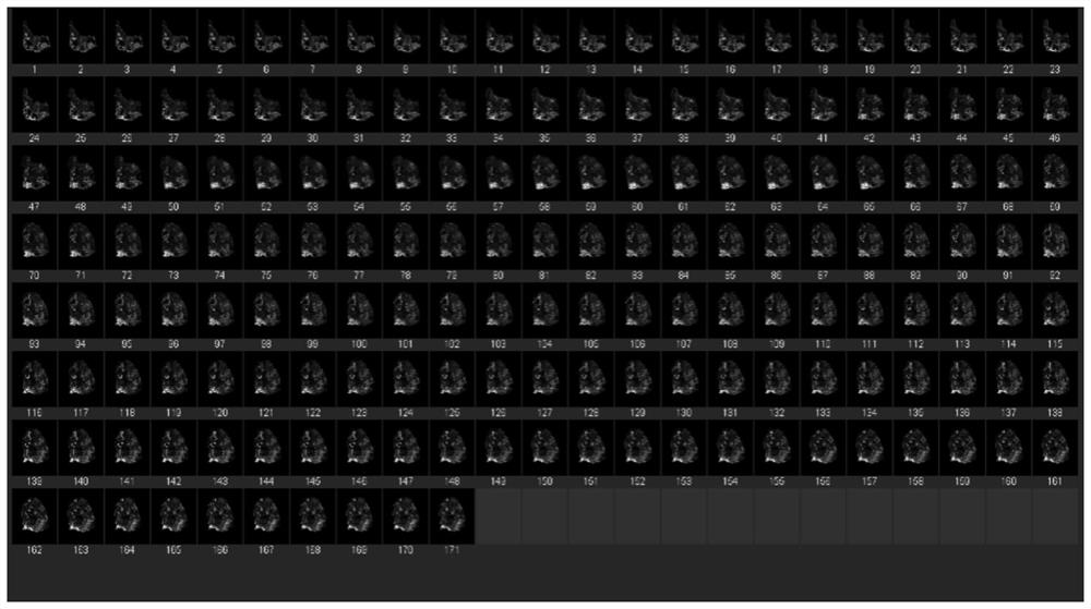
[0030]Embodiment 1: As shown in the figure, this three-dimensional imaging and analysis method of tissues and organs based on continuous slices, multicolor fluorescence and three-dimensional reconstruction mainly includes the following steps:

[0031]1) Take the reconstructed objects for strict serial sectioning; the reconstructed objects are isolated tissues and organs, which are frozen and paraffin-embedded for strict serial sectioning. Strictly continuous slicing. Each slice has a thickness of 2-10 microns, and each layer of slices is strictly marked with a serial number. For example, the first layer is marked as 1, the second layer is marked as 2, and so on. The section lost during the slicing process is also calculated in the marking Within the sequence.

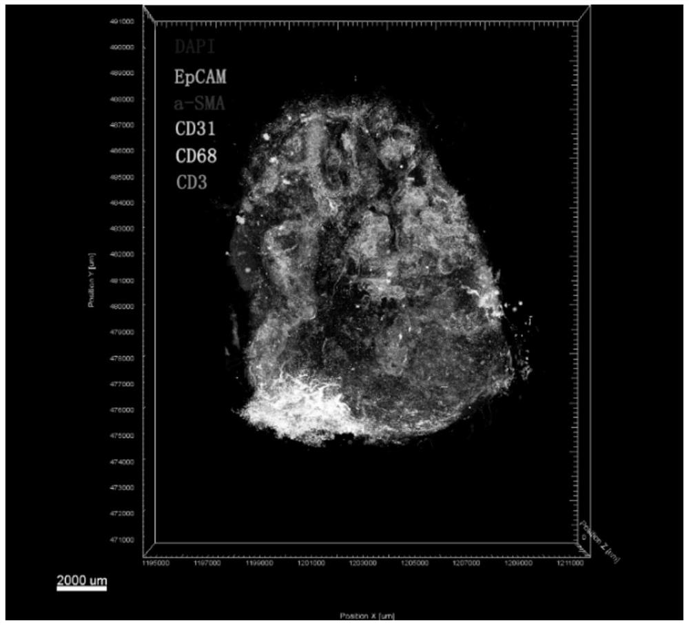
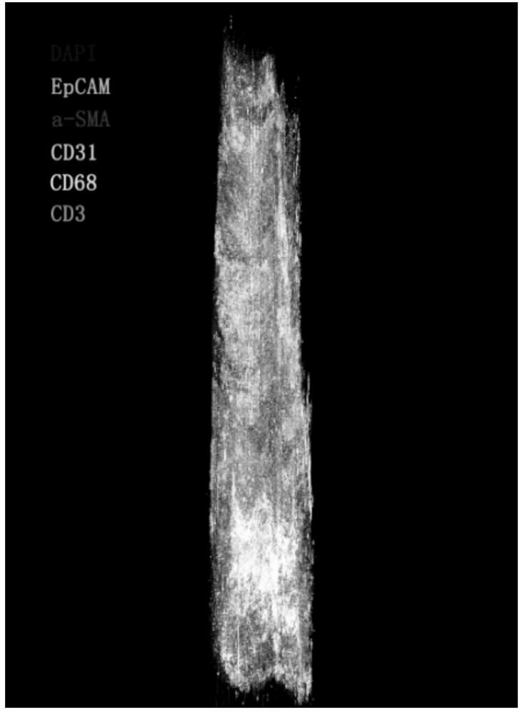
[0032]2) Multi-color immunofluorescence staining is performed alternately in multiple combinations according to the standard process; multi-color immunofluorescence staining uses multiple staining combinations to jointly participat...

Embodiment 2

[0036]Embodiment 2: This three-dimensional imaging and analysis method of tissues and organs based on serial slices, multi-color fluorescence and three-dimensional reconstruction, in specific implementation, mainly includes the following steps:

[0037]1) Tissue acquisition and preservation: take the isolated tissue 1cm*1cm*0.5cm for OCT freezing preservation or 10% formalin fixation for 48 hours;

[0038]2) Tissue treatment: If the tissue is fixed with 10% formalin, then dehydration: remove the tissue from the fixative solution in a fume hood, trim the tissue at the target site with a scalpel, and put the trimmed tissue into Corresponding to the numbered dehydration box. Put the dehydration box into the hanging basket and dehydrate with gradient alcohol in the dehydrator: 75% alcohol 2h, 85% alcohol 2h, 90% alcohol 2h, 95% alcohol 1h, absolute ethanol I1h, absolute ethanol II 1h, two Toluene I 45min, Xylene II 45min, Wax I1h-Wax II 1h, Wax III 1h. Followed by paraffin embedding: embed th...


PUM

PropertyMeasurementUnit
Thicknessaaaaaaaaaa

Abstract

The invention discloses a tissue and organ three-dimensional imaging and analysis method based on continuous slicing, multicolor fluorescence and three-dimensional reconstruction. The method mainly comprises the following steps: strictly and continuously slicing a reconstructed object; alternately carrying out multi-color immunofluorescence staining in multiple combinations according to a standardprocess; carrying out multi-channel fluorescence scanning on all the immunofluorescence staining sections to obtain digital section images; aligning continuous slices according to fluorescence channel signals, and inserting various dyeing combinations for three-dimensional reconstruction to obtain a tissue and organ fluorescence three-dimensional model with cell-level resolution; and conducting STL modeling on the interested local area, and conducting various kinds of analysis. According to the method, a high-resolution tissue and organ fluorescence three-dimensional model capable of performing quantitative analysis can be constructed, and the tissue and organ three-dimensional form can be faithfully restored to the maximum extent; one or more fluorescence channels can be checked independently or in an overlapping manner; and STL modeling can be performed on the region of interest to complete spatial quantification and correlation analysis of various cells and functional markers.

Description

Technical field[0001]The invention relates to the field of tissue and organ stereo imaging analysis, in particular to a tissue and organ stereo imaging and analysis method based on continuous slices, multicolor fluorescence and three-dimensional reconstruction.Background technique[0002]Tissue and organ specimens are important research materials in the field of biomedical research, as well as key specimens for disease diagnosis. Tissue imaging is an important means of diagnosis and research on tissue specimens. Tissue imaging mainly includes staining and detection.[0003]Staining methods include dye staining (such as HE staining) and antibody-based staining. The latter is divided into immunohistochemical staining (IHC) and immunofluorescence staining (IF). Among them, compared with IHC, IF can detect multiple antigens at the same time, which is helpful for more accurate cell identity confirmation and functional characterization.[0004]The detection part is still based on the observatio...

Claims


Application Information

Patent Timeline
IPC IPC(8): G01N21/64G01N15/14G01N1/30G01N1/28G01N33/53G01N33/533G01N33/58G06T17/00
CPCG01N1/28G01N1/30G01N15/1434G01N15/1468G01N21/6458G01N33/5302G01N33/533G01N33/582G01N2015/1445G01N2015/1472G06T17/00
Inventor 梁廷波章琦杨加琦白雪莉楼煜
Owner ZHEJIANG UNIV
Why Eureka
Try Eureka