DR-U-net network method and device for retinal blood flow image segmentation

A dr-u-net, image segmentation technology, applied in the field of DR-U-net network, to achieve the effect of accurate prediction model, increase network depth, and improve anti-interference performance

Pending Publication Date: 2021-02-09
FOSHAN UNIVERSITY
View PDF0 Cites 1 Cited by
  • Summary
  • Abstract
  • Description
  • Claims
  • Application Information

AI Technical Summary

Problems solved by technology

[0008] The purpose of the present invention is to propose a DR-U-net network method and device for retinal blood flow image segmentation, to solve one or more technical problems in the prior art, and at least provide a beneficial option or creating condition

Method used


Image

  • DR-U-net network method and device for retinal blood flow image segmentation
  • DR-U-net network method and device for retinal blood flow image segmentation

Examples


Embodiment Construction

[0045] The idea, specific structure and technical effects of the present invention will be clearly and completely described below in conjunction with the embodiments and accompanying drawings, so as to fully understand the purpose, scheme and effect of the present invention. It should be noted that, in the case of no conflict, the embodiments in the present application and the features in the embodiments can be combined with each other.

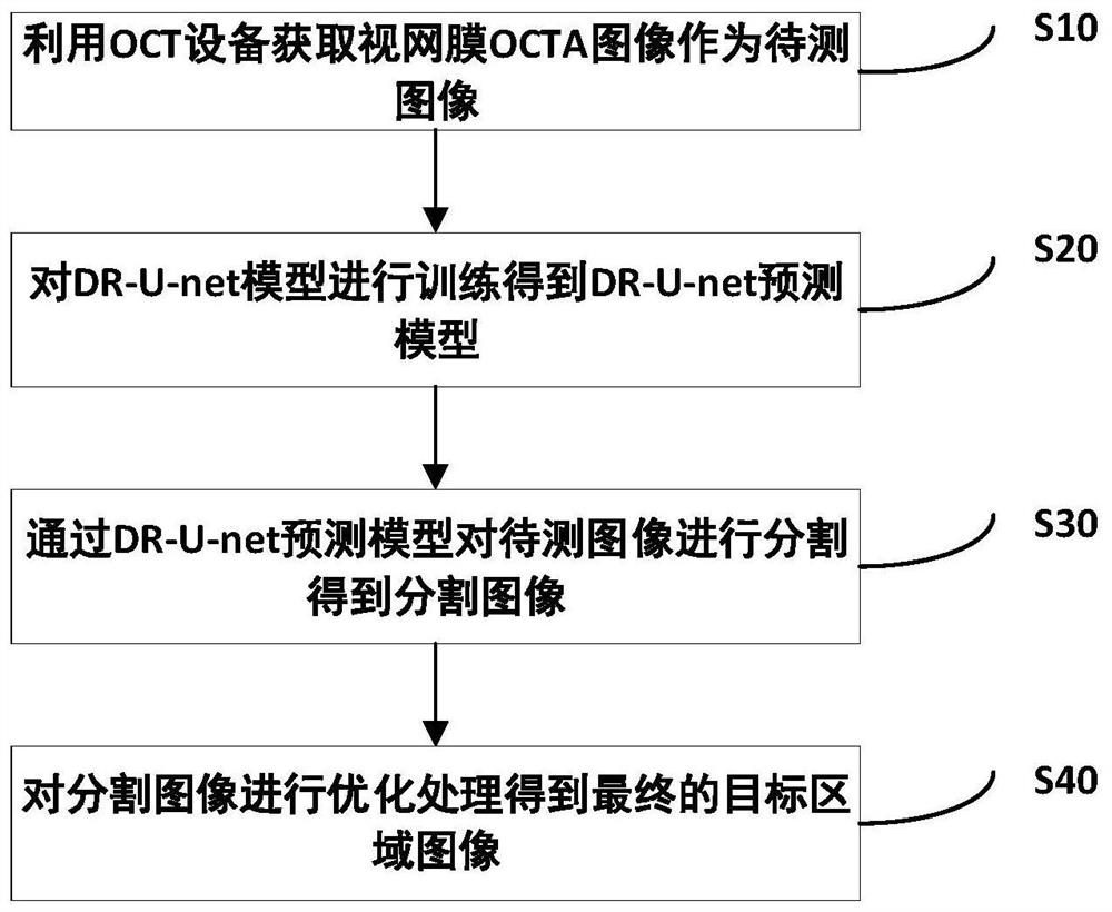
[0046] figure 1 Shown is the retinal OCTA image segmentation flow chart, combined below figure 1 A method according to an embodiment of the present invention will be described.

[0047] The present invention proposes a DR-U-net network method for retinal blood flow image segmentation, specifically comprising the following steps:

[0048] S10: Use the OCT device to obtain retinal OCTA images. The regions obtained by the OCT device are divided into non-retinal OCTA image regions, retinal OCTA image regions and background regions, and the reti...


PUM


Abstract

The invention discloses a DR-U-net network method and device for retinal blood flow image segmentation, and the method comprises the steps: carrying out the training of a DR-U-net prediction model, obtaining the DR-U-net prediction model, segmenting a to-be-detected image, carrying out the optimization of the segmented to-be-detected image, and obtaining a final target region; by combining a deeplearning method with a traditional image processing method, the problem of inaccurate segmentation easily generated when the signal-to-noise ratio of the retina OCTA image is low can be effectively solved, the segmentation precision of the retina OCTA image can be improved, and the anti-interference performance of the algorithm is improved. Fine features of the image can be acquired more effectively, so that a network model can acquire the fine features, and an optimal result can be obtained through segmentation; gradient disappearance or gradient explosion of the network in the network training process is avoided while the network depth is increased, feature multiplexing can be more effectively carried out, and the prediction model is more accurate.

Description

technical field [0001] The invention relates to the field of optical coherence tomography, in particular to a DR-U-net network method and device for retinal blood flow image segmentation. Background technique [0002] Optical coherence tomography (OCT) is a new three-dimensional tomography technology developed gradually in the 1990s. Due to the continuous maturity of OCT technology, it has become the gold standard for the diagnosis and evaluation of ophthalmic diseases. In recent years, the rapid development of OCT has also led to the development of optical coherence tomography angiography (OCTA). OCTA uses low-coherence interferometry to measure changes in the backscatter signal to distinguish areas of blood flow from areas of static tissue, and repeatedly measures the phase and intensity of retinal microvessels at the same scan location to measure blood flow in retinal microvessels. OCTA can visualize the superficial and deep capillaries of retinal vessels, and it does n...

Claims


Application Information

Patent Timeline
Patent Type & Authority Applications(China)
IPC IPC(8): G06T7/11G06T7/13G06N3/04G06N3/08G06K9/62
CPCG06T7/11G06T7/13G06N3/08G06T2207/30041G06T2207/20021G06N3/044G06F18/214
Inventor 袁钘许景江韦赢兆安林黄燕平蓝公仆秦嘉
Owner FOSHAN UNIVERSITY
Why Eureka
Try Eureka