Animal tracheal cannula device

A technology for tracheal intubation and animals, which is applied in the direction of tracheal intubation and respirators, etc., which can solve the problems of large tracheal damage and easily blocked vision, and achieve the effects of improved accuracy, less damage, and compact structure

Active Publication Date: 2021-02-19
RWD LIFE SCI
View PDF7 Cites 0 Cited by
  • Summary
  • Abstract
  • Description
  • Claims
  • Application Information

AI Technical Summary

Problems solved by technology

[0003] The embodiment of the present invention provides an animal tracheal intubation device, which aims to solve the problems in

Method used


Image

  • Animal tracheal cannula device
  • Animal tracheal cannula device

Examples


Embodiment 1

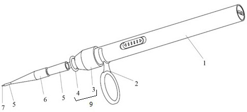
[0027] figure 1 It is a schematic diagram of the animal tracheal intubation device provided in Embodiment 1 of the present invention. Such as figure 1 As shown, the animal tracheal intubation device includes an LED light source 1 , a connecting part 9 , a tracheal intubation tube 5 and an optical fiber 7 .

[0028] In the embodiment of the present invention, the LED light source 1 is a wireless power supply, adopts a USB fast charging design, can be used without a power cord, and increases the portability and movable range of the device; when the light intensity is insufficient, the device can be charged, After charging is complete, you can unplug the power cord and continue to use it. The light intensity of the LED light source 1 is adjustable. Preferably, it is divided into two levels of strong and weak, which can be switched according to needs, so as to prevent users from working under strong light for a long time to affect their eyesight. Using the above-mentioned LED l...

Embodiment 2

[0035] Such as figure 1 As shown, the connection part 9 includes a first connection part 3 and a second connection part 4 . Both the first connecting part 3 and the second connecting part 4 are hollow. The first connecting part 3 is sleeved on the light-emitting end of the LED light source 1, the second connecting part 4 is connected with the other end of the first connecting part 3 with a taper, and the endotracheal tube 5 is also connected with the other end of the second connecting part 4 with a taper. The optical fiber 7 passes through the second connection part 4 and is locked therein.

[0036] As another embodiment of the present invention, in order to adapt to the size of the trachea of ​​different animals, preferably, the second connecting part 4 is provided with silica gel inside to lock the optical fiber 7 . Users can select optical fibers 7 with different diameters according to their needs, and fix them through the silica gel. The first end of the optical fiber 7...

Embodiment 3

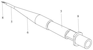
[0040] figure 2 It is a schematic diagram of the animal tracheal intubation device provided in Embodiment 3 of the present invention. In the embodiment of the present invention, the endotracheal tube 5 and the second connecting part 4 are detachable. After successful intubation, the LED light source 1, the magnifying glass 2, the first connecting part 3, the second connecting part 4 and the optical fiber 7 are removed, and the tracheal intubation tube 5 and the safety plug 6 are left in the trachea of ​​the animal. The cannula 5 can be connected to a ventilator or administered intratracheally through a drug delivery needle.

[0041] Such as figure 1 and figure 2 As shown, the animal endotracheal intubation device includes an LED light source 1 , a magnifying glass 2 , a first connection part 3 , a second connection part 4 , an endotracheal tube 5 , a safety plug 6 and an optical fiber 7 , and also includes a drug delivery needle 8 . After the second connecting part 4 is ...


PUM


Abstract

The embodiment of the invention provides an animal trachea cannula device. The animal trachea cannula device comprises an LED light source (1), a connecting component (9) connected to the light-emitting end of the LED light source (1) in a sleeving mode, an optical fiber (7) which passes through the connection component (9) and is locked, and an trachea cannula (5) in taper connection with the connecting component (9), wherein the first end of the optical fiber (7) is connected with the light-emitting end of the LED light source (1), and the second end of the optical fiber (7) penetrates through the trachea cannula (5). According to the trachea cannula, light of the LED light source is guided out to the front end of the trachea cannula through the optical fiber, the connecting component and the trachea cannula are in taper connection, the trachea cannula can be matched with trachea cannulas of different specifications, the structure is compact, handheld operation is convenient, the LEDlight source cannot be shielded when the trachea cannula is used for intubation of animals, intubation accuracy is improved, and damage to the trachea is small.

Description

technical field [0001] The invention relates to the technical field of animal medical instruments, in particular to an animal trachea intubation device. Background technique [0002] Currently, there are two main methods for non-invasive intubation in animals: blind intubation and intubation under a laryngoscope. Due to the low degree of visibility, the blind insertion method is easy to damage the epiglottis of experimental animals, and it takes a long time, the success rate is low, it is easy to cause infection, and the wound is not easy to recover. On the contrary, it increases the pathological state of the animal and easily leads to death of the animal. affect the experiment process. When intubating under a laryngoscope, although the laryngoscope is used as a lighting tool, the endotracheal intubation often blocks the light source when passing through the epiglottis, which reduces the accuracy of intubation, and the operation takes a long time. Not only that, when intub...

Claims


Application Information

Patent Timeline
IPC IPC(8): A61M16/04
CPCA61M16/04A61M16/0488
Inventor 郭俊秀陈福县罗哲民
Owner RWD LIFE SCI
Why Eureka
Try Eureka